Deposition Date
2011-04-07
Release Date
2011-11-23
Last Version Date
2024-05-08
Entry Detail
PDB ID:
2YFQ
Keywords:
Title:
Crystal structure of Glutamate dehydrogenase from Peptoniphilus asaccharolyticus
Biological Source:
Source Organism(s):
PEPTONIPHILUS ASACCHAROLYTICUS (Taxon ID: 1258)
Expression System(s):
Method Details:
Experimental Method:
Resolution:
2.94 Å
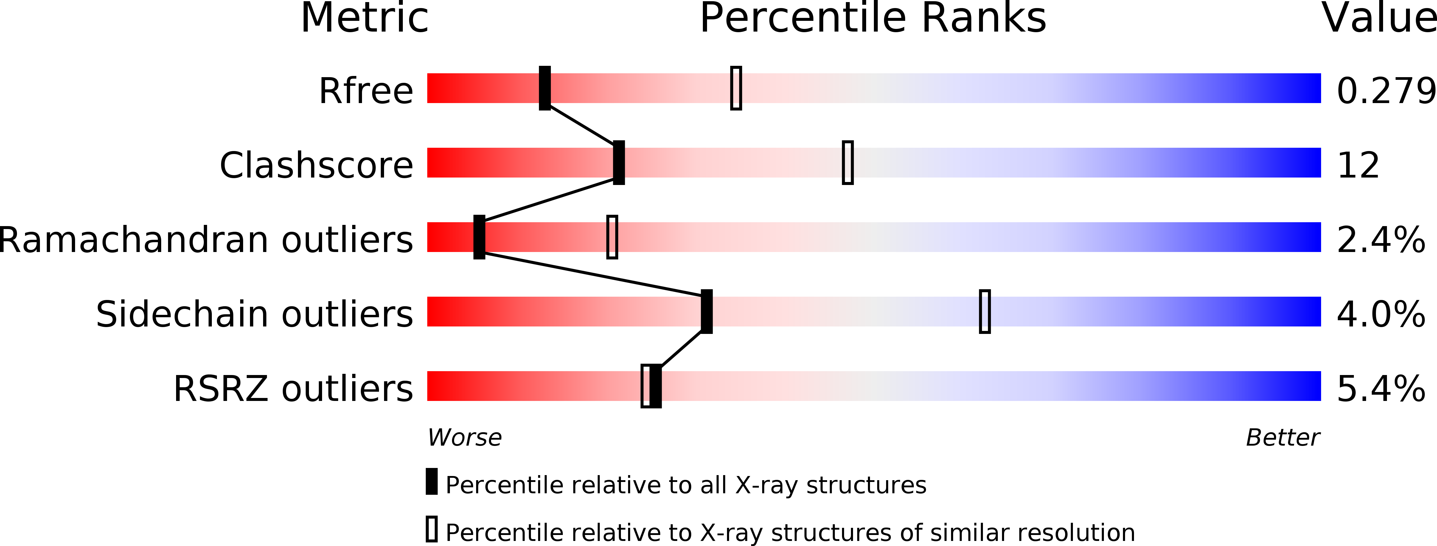
R-Value Free:
0.28
R-Value Work:
0.24
R-Value Observed:
0.24
Space Group:
H 3 2