Deposition Date
2011-03-28
Release Date
2012-02-08
Last Version Date
2024-11-06
Entry Detail
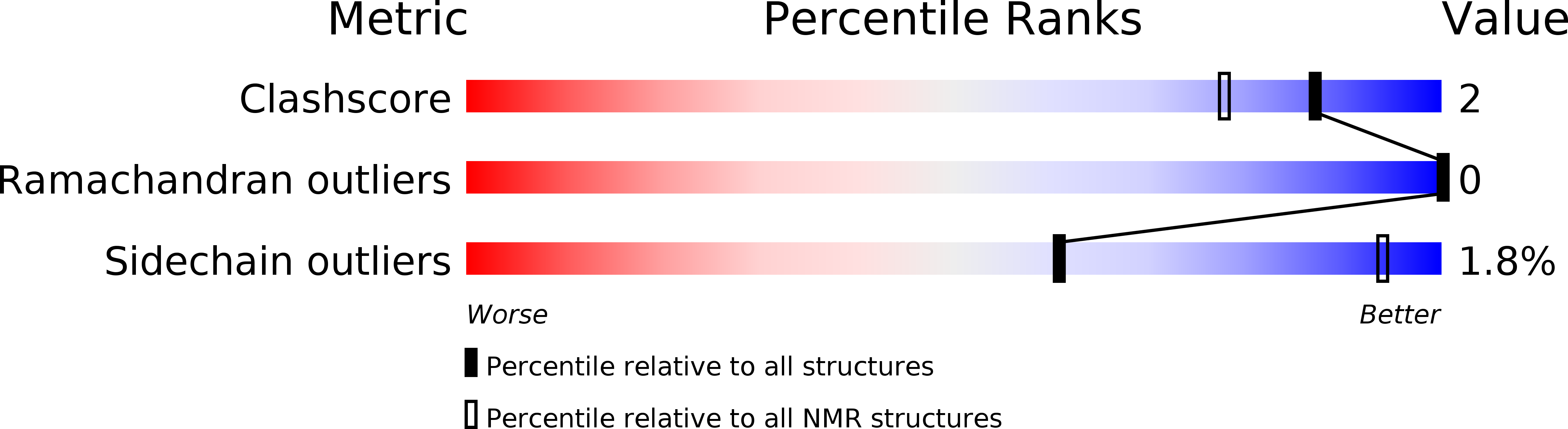
PDB ID:
2YEN
Keywords:
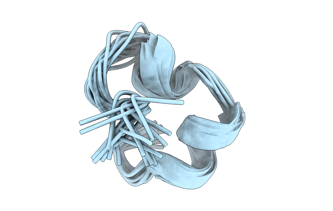
Title:
Solution structure of the skeletal muscle and neuronal voltage gated sodium channel antagonist mu-conotoxin CnIIIC
Biological Source:
Source Organism(s):
Conus consors (Taxon ID: 101297)
Method Details:
Experimental Method:
Conformers Calculated:
200
Conformers Submitted:
20
Selection Criteria:
LEAST RESTRAINT VIOLATION