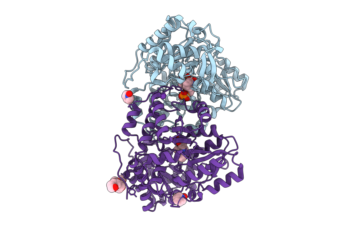
2YCT image
Entry Detail
PDB ID:
2YCT
Keywords:
Title:
Tyrosine phenol-lyase from Citrobacter freundii in complex with pyridine N-oxide and the quinonoid intermediate formed with L-alanine
Biological Source:
Source Organism:
Host Organism:
PDB Version:
Deposition Date:
2011-03-16
Release Date:
2011-09-14
Method Details:
Experimental Method:
Resolution:
2.25 Å
R-Value Free:
0.18
R-Value Work:
0.13
R-Value Observed:
0.14
Space Group:
P 21 21 2
Macromolecular Entities
Polymer Type:polypeptide(L)
Description:TYROSINE PHENOL-LYASE
Chain IDs:A, B
Chain Length:456
Number of Molecules:2
Biological Source:CITROBACTER FREUNDII
Primary Citation
Crystallographic Snapshots of Tyrosine Phenol-Lyase Show that Substrate Strain Plays a Role in C-C Bond Cleavage
J.Am.Chem.Soc. 133 16468 ? (2011)
PMID: 21899319 DOI: 10.1021/JA203361G

Abstact

The key step in the enzymatic reaction catalyzed by tyrosine phenol-lyase (TPL) is reversible cleavage of the Cβ-Cγ bond of L-tyrosine. Here, we present X-ray structures for two enzymatic states that form just before and after the cleavage of the carbon-carbon bond. As for most other pyridoxal 5'-phosphate-dependent enzymes, the first state, a quinonoid intermediate, is central for the catalysis. We captured this relatively unstable intermediate in the crystalline state by introducing substitutions Y71F or F448H in Citrobacter freundii TPL and briefly soaking crystals of the mutant enzymes with a substrate 3-fluoro-L-tyrosine followed by flash-cooling. The X-ray structures, determined at ~2.0 Å resolution, reveal two quinonoid geometries: "relaxed" in the open and "tense" in the closed state of the active site. The "tense" state is characterized by changes in enzyme contacts made with the substrate's phenolic moiety, which result in significantly strained conformation at Cβ and Cγ positions. We also captured, at 2.25 Å resolution, the X-ray structure for the state just after the substrate's Cβ-Cγ bond cleavage by preparing the ternary complex between TPL, alanine quinonoid and pyridine N-oxide, which mimics the α-aminoacrylate intermediate with bound phenol. In this state, the enzyme-ligand contacts remain almost exactly the same as in the "tense" quinonoid, indicating that the strain induced by the closure of the active site facilitates elimination of phenol. Taken together, structural observations demonstrate that the enzyme serves not only to stabilize the transition state but also to destabilize the ground state.

Legend

Protein

Chemical

Disease

Primary Citation of related structures