Deposition Date
2011-02-23
Release Date
2011-06-08
Last Version Date
2023-12-20
Entry Detail
PDB ID:
2YAL
Keywords:
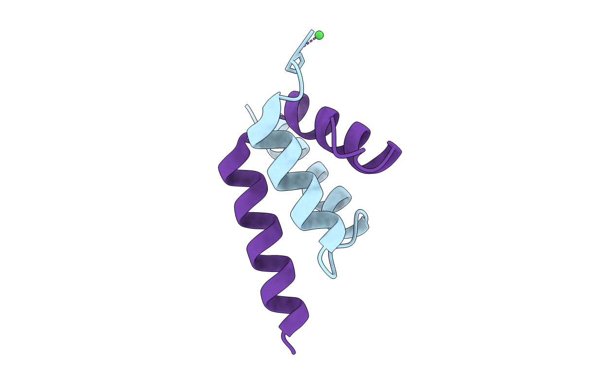
Title:
SinR, Master Regulator of biofilm formation in Bacillus subtilis
Biological Source:
Source Organism(s):
BACILLUS SUBTILIS (Taxon ID: 1423)
Expression System(s):
Method Details:
Experimental Method:
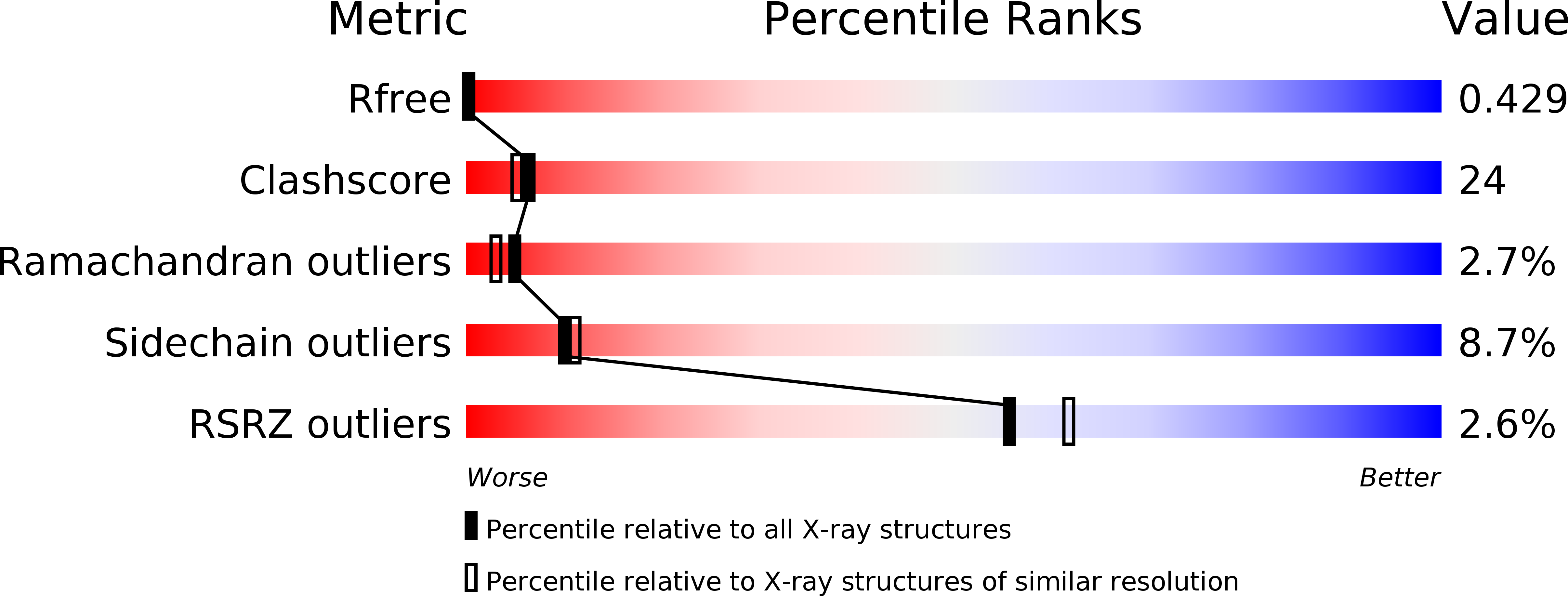
Resolution:
2.27 Å
R-Value Free:
0.43
R-Value Work:
0.28
R-Value Observed:
0.28
Space Group:
P 61 2 2