Deposition Date
2011-01-27
Release Date
2011-09-28
Last Version Date
2023-12-20
Entry Detail
PDB ID:
2Y72
Keywords:
Title:
Crystal structure of the PKD Domain of Collagenase G from Clostridium Histolyticum at 1.18 Angstrom Resolution.
Biological Source:
Source Organism(s):
CLOSTRIDIUM HISTOLYTICUM (Taxon ID: 1498)
Expression System(s):
Method Details:
Experimental Method:
Resolution:
1.18 Å
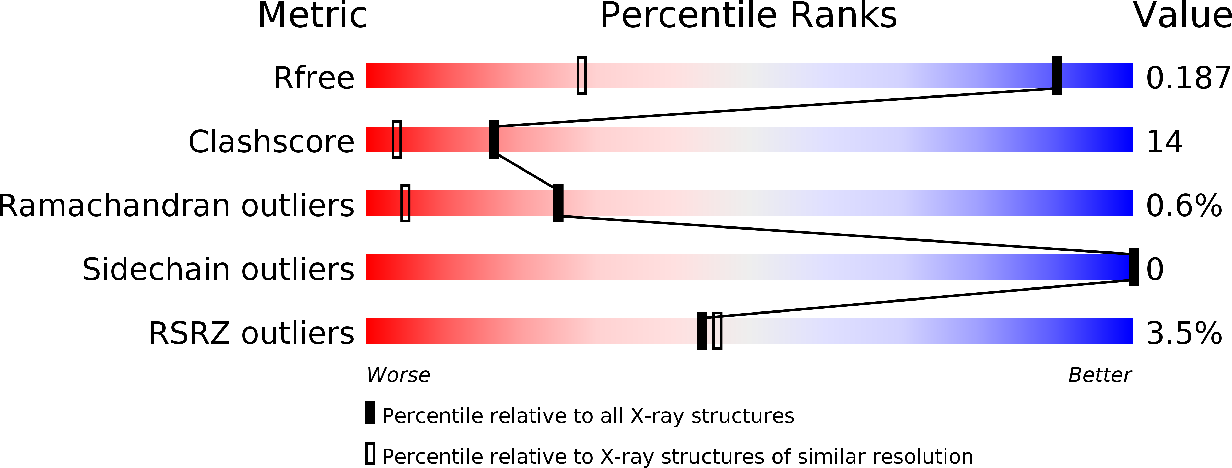
R-Value Free:
0.17
R-Value Work:
0.14
R-Value Observed:
0.14
Space Group:
C 1 2 1