2Y6X image
Deposition Date 2011-01-27
Release Date 2012-01-11
Last Version Date 2023-12-20
Entry Detail
PDB ID:
2Y6X
Keywords:
Title:
Structure of Psb27 from Thermosynechococcus elongatus
Biological Source:
Source Organism:
Host Organism:
Method Details:
Experimental Method:
Resolution:
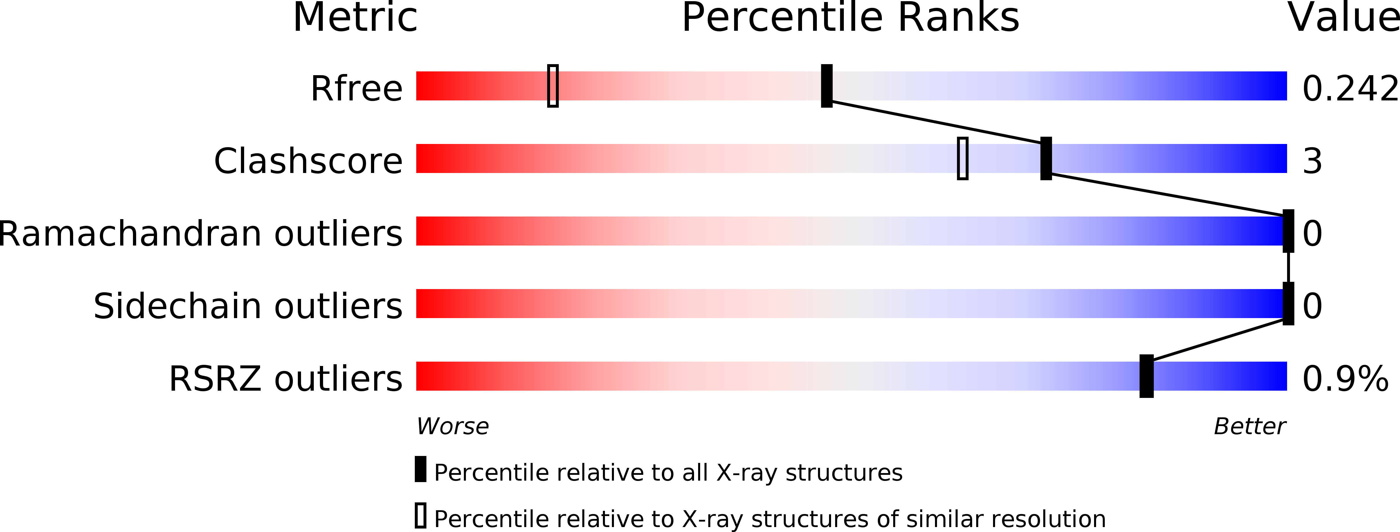
1.60 Å
R-Value Free:
0.23
R-Value Work:
0.19
R-Value Observed:
0.19
Space Group:
P 21 21 21
Macromolecular Entities
Polymer Type:polypeptide(L)
Molecule:PHOTOSYSTEM II 11 KD PROTEIN
Gene (Uniprot):psb27
Chain IDs:A
Chain Length:113
Number of Molecules:1
Biological Source:THERMOSYNECHOCOCCUS ELONGATUS
Ligand Molecules
Primary Citation
Crystal Structure of the Psb27 Assembly Factor at 1.6A: Implications for Binding to Photosystem II.
Photosynth.Res. 110 169 ? (2012)
PMID: 22193820 DOI: 10.1007/S11120-011-9712-7

Abstact

The biogenesis and oxygen-evolving activity of cyanobacterial Photosystem II (PSII) is dependent on a number of accessory proteins not found in the crystallised dimeric complex. These include Psb27, a small lipoprotein attached to the lumenal side of PSII, which has been assigned a role in regulating the assembly of the Mn(4)Ca cluster catalysing water oxidation. To gain a better understanding of Psb27, we have determined in this study the crystal structure of the soluble domain of Psb27 from Thermosynechococcus elongatus to a resolution of 1.6 Å. The structure is a four-helix bundle, similar to the recently published solution structures of Psb27 from Synechocystis PCC 6803 obtained by nuclear magnetic resonance (NMR) spectroscopy. Importantly, the crystal structure presented here helps us resolve the differences between the NMR-derived structural models. Potential binding sites for Psb27 within PSII are discussed in light of recent biochemical data in the literature.

Legend

Protein

Chemical

Disease

Primary Citation of related structures