2Y6S image
Deposition Date 2011-01-25
Release Date 2012-01-11
Last Version Date 2024-10-23
Entry Detail
PDB ID:
2Y6S
Title:
Structure of an Ebolavirus-protective antibody in complex with its mucin-domain linear epitope
Biological Source:
Source Organism:
ZAIRE EBOLAVIRUS (Taxon ID: 128951)
MUS MUSCULUS (Taxon ID: 10090)
Method Details:
Experimental Method:
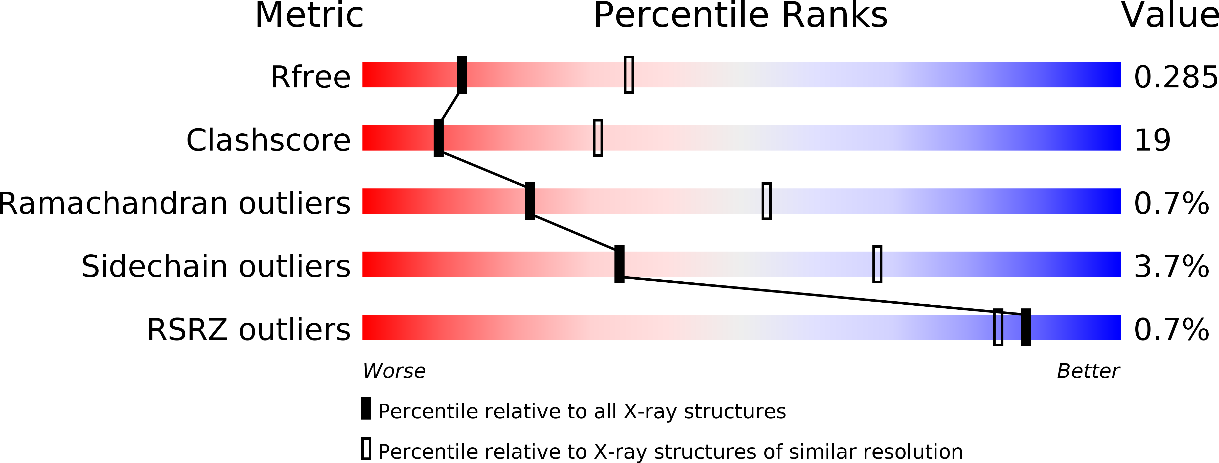
Resolution:
2.80 Å
R-Value Free:
0.28
R-Value Work:
0.23
R-Value Observed:
0.23
Space Group:
P 1 2 1
Macromolecular Entities
Polymer Type:polypeptide(L)
Molecule:LIGHT CHAIN
Chain IDs:A (auth: C), D (auth: L)
Chain Length:217
Number of Molecules:2
Biological Source:MUS MUSCULUS
Polymer Type:polypeptide(L)
Molecule:HEAVY CHAIN
Chain IDs:B (auth: D), C (auth: H)
Chain Length:213
Number of Molecules:2
Biological Source:MUS MUSCULUS
Polymer Type:polypeptide(L)
Molecule:ENVELOPE GLYCOPROTEIN
Gene (Uniprot):GP
Chain IDs:E (auth: P), F (auth: Q)
Chain Length:17
Number of Molecules:2
Biological Source:ZAIRE EBOLAVIRUS
Primary Citation
Structure of an Ebola Virus-Protective Antibody in Complex with its Mucin-Domain Linear Epitope.
J.Virol. 86 2809 ? (2012)
PMID: 22171276 DOI: 10.1128/JVI.05549-11

Abstact

Antibody 14G7 is protective against lethal Ebola virus challenge and recognizes a distinct linear epitope in the prominent mucin-like domain of the Ebola virus glycoprotein GP. The structure of 14G7 in complex with its linear peptide epitope has now been determined to 2.8 Å. The structure shows that this GP sequence forms a tandem β-hairpin structure that binds deeply into a cleft in the antibody-combining site. A key threonine at the apex of one turn is critical for antibody interaction and is conserved among all Ebola viruses. This work provides further insight into the mechanism of protection by antibodies that target the protruding, highly accessible mucin-like domain of Ebola virus and the structural framework for understanding and characterizing candidate immunotherapeutics.

Legend

Protein

Chemical

Disease

Primary Citation of related structures