2Y3U image
Deposition Date 2010-12-26
Release Date 2011-09-28
Last Version Date 2024-11-20
Entry Detail
PDB ID:
2Y3U
Keywords:
Title:
Crystal structure of apo collagenase G from Clostridium histolyticum at 2.55 Angstrom resolution
Biological Source:
Source Organism:
Host Organism:
Method Details:
Experimental Method:
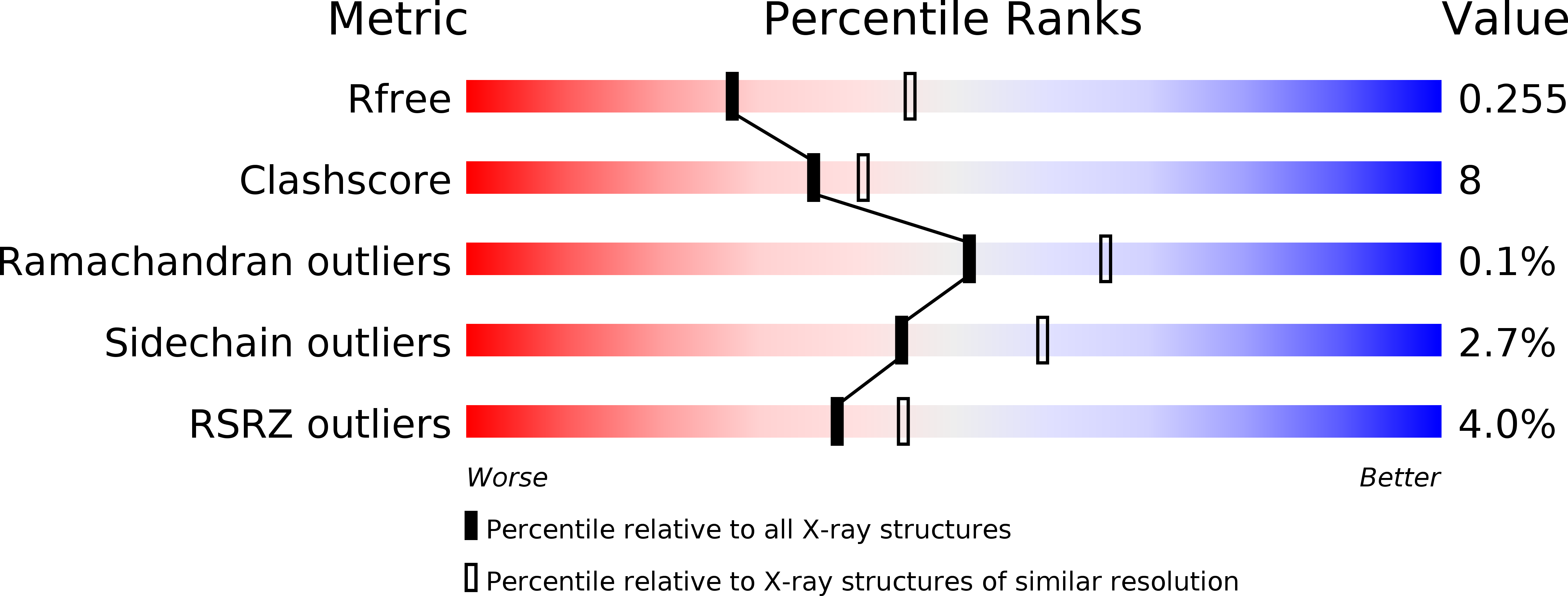
Resolution:
2.55 Å
R-Value Free:
0.25
R-Value Work:
0.20
R-Value Observed:
0.21
Space Group:
P 21 21 21
Macromolecular Entities
Polymer Type:polypeptide(L)
Molecule:COLLAGENASE
Gene (Uniprot):colG
Chain IDs:A
Chain Length:785
Number of Molecules:1
Biological Source:CLOSTRIDIUM HISTOLYTICUM
Modified Residue
Compound ID Chain ID Parent Comp ID Details 2D Image
MSE A MET SELENOMETHIONINE
Primary Citation
Structure of Collagenase G Reveals a Chew-and -Digest Mechanism of Bacterial Collagenolysis
Nat.Struct.Mol.Biol. 18 1109 ? (2011)
PMID: 21947205 DOI: 10.1038/NSMB.2127

Abstact

Collagen constitutes one-third of body protein in humans, reflecting its extensive role in health and disease. Of similar importance, therefore, are the idiosyncratic proteases that have evolved for collagen remodeling. The most efficient collagenases are those that enable clostridial bacteria to colonize their host tissues; but despite intense study, the structural and mechanistic basis of these enzymes has remained elusive. Here we present the crystal structure of collagenase G from Clostridium histolyticum at 2.55-Å resolution. By combining the structural data with enzymatic and mutagenesis studies, we derive a conformational two-state model of bacterial collagenolysis, in which recognition and unraveling of collagen microfibrils into triple helices, as well as unwinding of the triple helices, are driven by collagenase opening and closing.

Legend

Protein

Chemical

Disease

Primary Citation of related structures