Deposition Date
2010-12-22
Release Date
2010-12-29
Last Version Date
2023-12-20
Entry Detail
PDB ID:
2Y3P
Keywords:
Title:
Crystal structure of N-terminal domain of GyrA with the antibiotic simocyclinone D8
Biological Source:
Source Organism(s):
ESCHERICHIA COLI (Taxon ID: 562)
Expression System(s):
Method Details:
Experimental Method:
Resolution:
2.62 Å
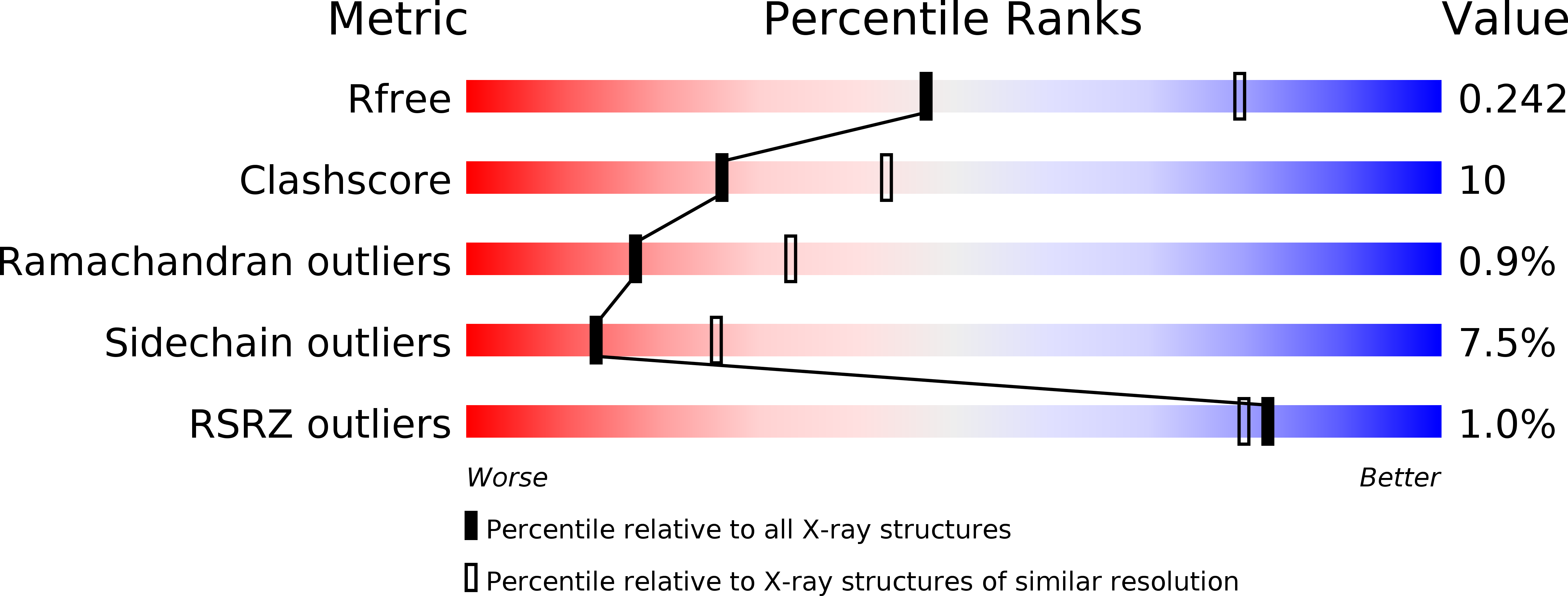
R-Value Free:
0.24
R-Value Work:
0.20
R-Value Observed:
0.20
Space Group:
I 21 21 21