Deposition Date
2010-12-21
Release Date
2011-11-02
Last Version Date
2024-05-08
Entry Detail
PDB ID:
2Y3L
Keywords:
Title:
Structure of segment MVGGVVIA from the amyloid-beta peptide (Ab, residues 35-42), alternate polymorph 2
Biological Source:
Source Organism(s):
HOMO SAPIENS (Taxon ID: 9606)
Method Details:
Experimental Method:
Resolution:
2.10 Å
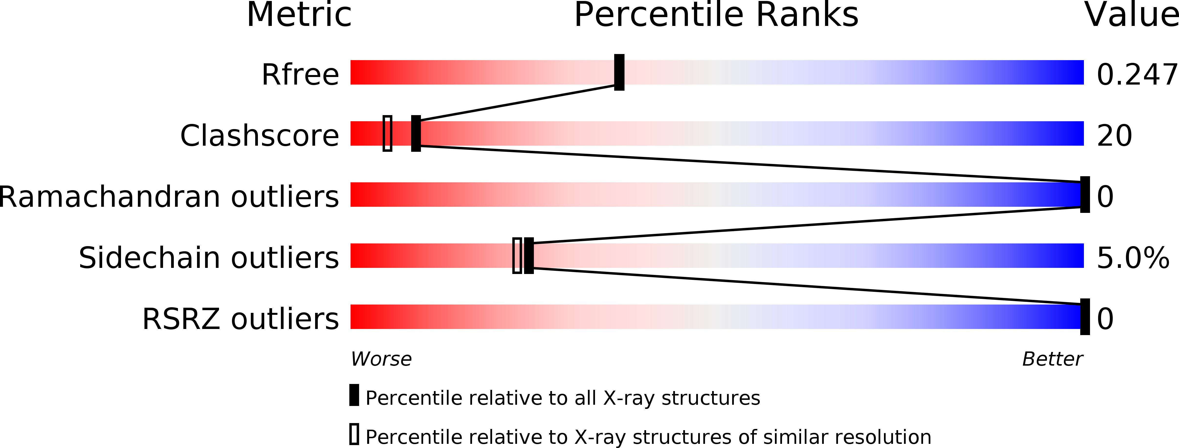
R-Value Free:
0.24
R-Value Work:
0.20
R-Value Observed:
0.20
Space Group:
P 1 21 1