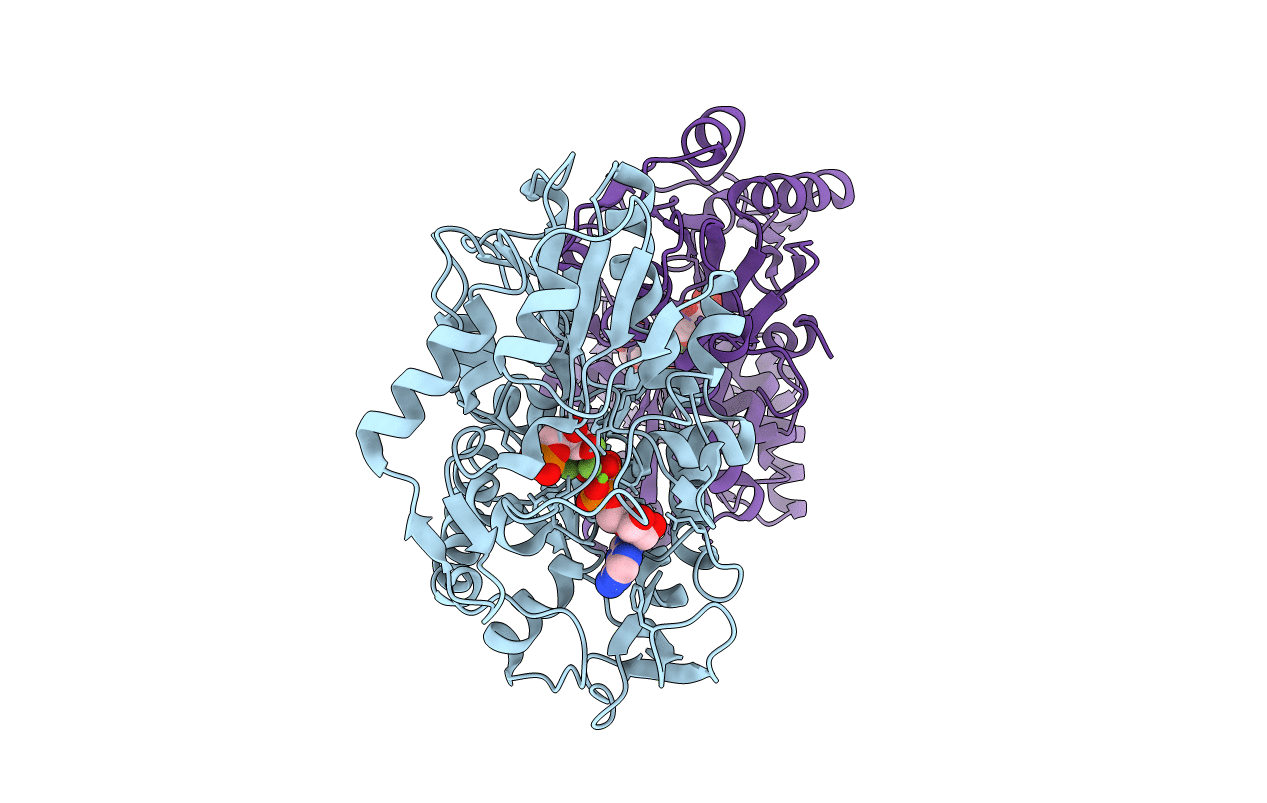
2Y3I image
Entry Detail
PDB ID:
2Y3I
Keywords:
Title:
The structure of the fully closed conformation of human PGK in complex with L-ADP, 3PG and the TSA aluminium tetrafluoride
Biological Source:
Source Organism:
Host Organism:
PDB Version:
Deposition Date:
2010-12-21
Release Date:
2011-04-27
Method Details:
Experimental Method:
Resolution:
2.90 Å
R-Value Free:
0.30
R-Value Work:
0.26
R-Value Observed:
0.26
Space Group:
P 21 21 21
Macromolecular Entities
Polymer Type:polypeptide(L)
Description:PHOSPHOGLYCERATE KINASE 1
Chain IDs:A, B (auth: D)
Chain Length:416
Number of Molecules:2
Biological Source:HOMO SAPIENS
Primary Citation
Interaction of Human 3-Phosphoglycerate Kinase with its Two Substrates: Is Substrate Antagonism a Kinetic Advantage?
J.Mol.Biol. 409 742 ? (2011)
PMID: 21549713 DOI: 10.1016/J.JMB.2011.04.048

Abstact

Substrate antagonism has been described for a variety of enzymes with more than one substrate and is characterized by a lowering of the affinity of one substrate in the presence of the other(s). 3-Phosphoglycerate kinase (PGK) catalyzes phosphotransfer from 1,3-bisphosphoglycerate (bPG) to ADP to give 3-phosphoglycerate (PG) and ATP, and is subject to substrate antagonism. Because of the instability of bPG, antagonism has only been described between PG and ATP or ADP. Here, we show that antagonism also occurs between bPG and ADP. Using the stopped-flow method, we show that the dissociation constant for one substrate increases in the presence of the other, and that this decrease in affinity is mainly due to an increase in the dissociation rate constant. As a consequence, there is an increase in the overall interaction kinetics. Interestingly, in the presence of the mirror image of natural d-ADP, l-ADP (a good substrate for PGK), antagonism is absent. Using rapid-quench-flow, we studied the kinetics of ATP formation. The time courses present the following: (1) a lag with l-ADP, but not with d-ADP, the kinetics of which were similar to the interaction kinetics measured by stopped-flow; (2) a burst that is directed by the phosphotransfer; and (3) a steady-state that is rate limited by the release of product kinetics. Structural explanations for these results are proposed by analyzing the crystallographic structure of the fully closed conformation of PGK in complex with l-ADP, PG, and the transition-state analogue AlF(4)(-) compared to previously determined structures.

Legend

Protein

Chemical

Disease

Primary Citation of related structures