2Y2B image
Deposition Date 2010-12-14
Release Date 2011-07-20
Last Version Date 2024-01-31
Entry Detail
PDB ID:
2Y2B
Keywords:
Title:
crystal structure of AmpD in complex with reaction products
Biological Source:
Source Organism:
Host Organism:
Method Details:
Experimental Method:
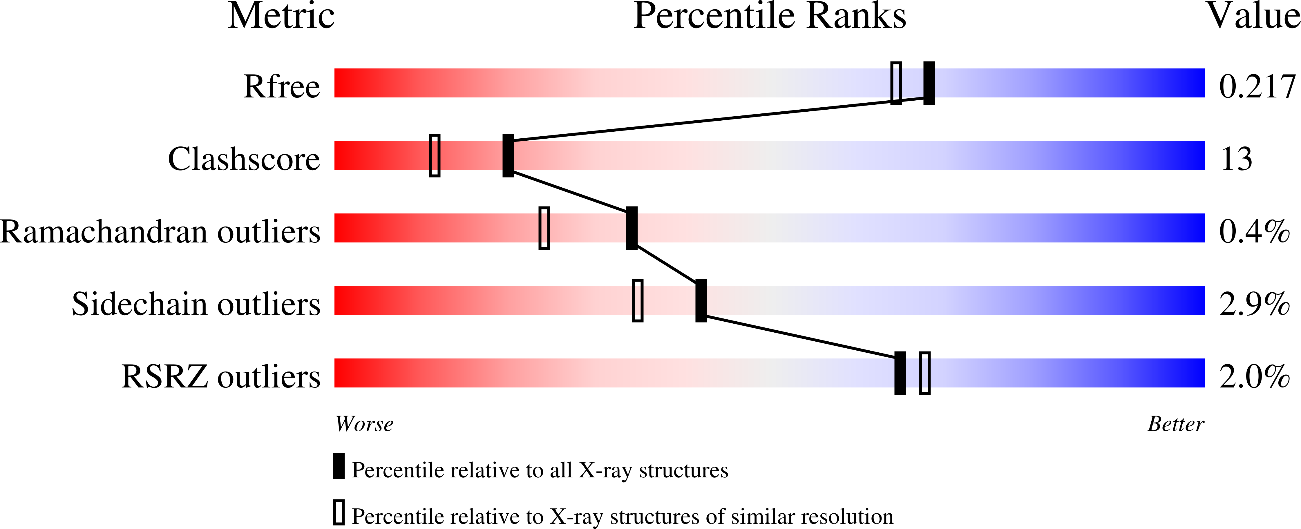
Resolution:
1.90 Å
R-Value Free:
0.22
R-Value Work:
0.16
R-Value Observed:
0.17
Space Group:
P 32
Macromolecular Entities
Polymer Type:polypeptide(L)
Molecule:1,6-ANHYDRO-N-ACETYLMURAMYL-L-ALANINE AMIDASE AMPD
Gene (Uniprot):ampD
Chain IDs:A, B, C
Chain Length:187
Number of Molecules:3
Biological Source:CITROBACTER FREUNDII
Primary Citation
Crystal Structures of Bacterial Peptidoglycan Amidase Ampd and an Unprecedented Activation Mechanism.
J.Biol.Chem. 286 31714 ? (2011)
PMID: 21775432 DOI: 10.1074/JBC.M111.264366

Abstact

AmpD is a cytoplasmic peptidoglycan (PG) amidase involved in bacterial cell-wall recycling and in induction of β-lactamase, a key enzyme of β-lactam antibiotic resistance. AmpD belongs to the amidase_2 family that includes zinc-dependent amidases and the peptidoglycan-recognition proteins (PGRPs), highly conserved pattern-recognition molecules of the immune system. Crystal structures of Citrobacter freundii AmpD were solved in this study for the apoenzyme, for the holoenzyme at two different pH values, and for the complex with the reaction products, providing insights into the PG recognition and the catalytic process. These structures are significantly different compared with the previously reported NMR structure for the same protein. The NMR structure does not possess an accessible active site and shows the protein in what is proposed herein as an inactive "closed" conformation. The transition of the protein from this inactive conformation to the active "open" conformation, as seen in the x-ray structures, was studied by targeted molecular dynamics simulations, which revealed large conformational rearrangements (as much as 17 Å) in four specific regions representing one-third of the entire protein. It is proposed that the large conformational change that would take the inactive NMR structure to the active x-ray structure represents an unprecedented mechanism for activation of AmpD. Analysis is presented to argue that this activation mechanism might be representative of a regulatory process for other intracellular members of the bacterial amidase_2 family of enzymes.

Legend

Protein

Chemical

Disease

Primary Citation of related structures