Deposition Date
2010-12-10
Release Date
2011-11-09
Last Version Date
2024-10-16
Entry Detail
PDB ID:
2Y1V
Keywords:
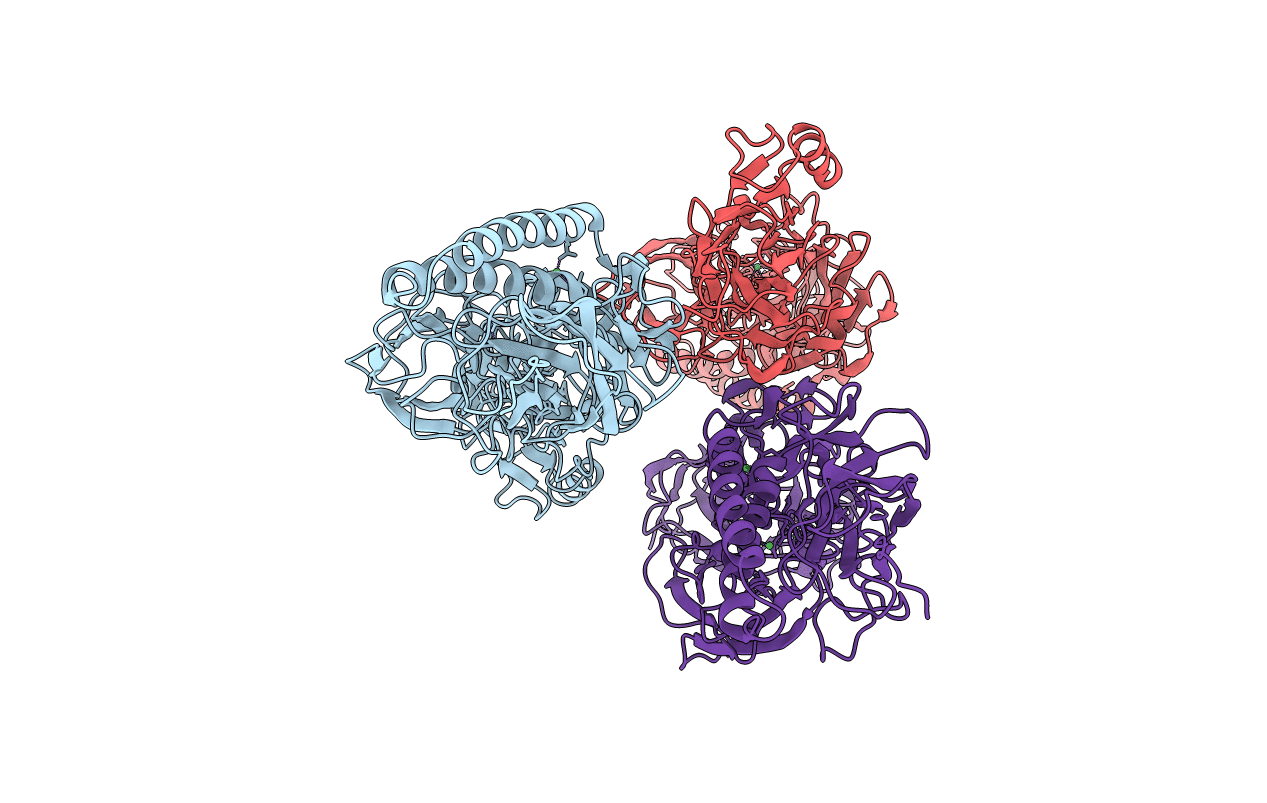
Title:
Full length structure of RrgB Pilus protein from Streptococcus pneumoniae
Biological Source:
Source Organism(s):
STREPTOCOCCUS PNEUMONIAE (Taxon ID: 170187)
Expression System(s):
Method Details:
Experimental Method:
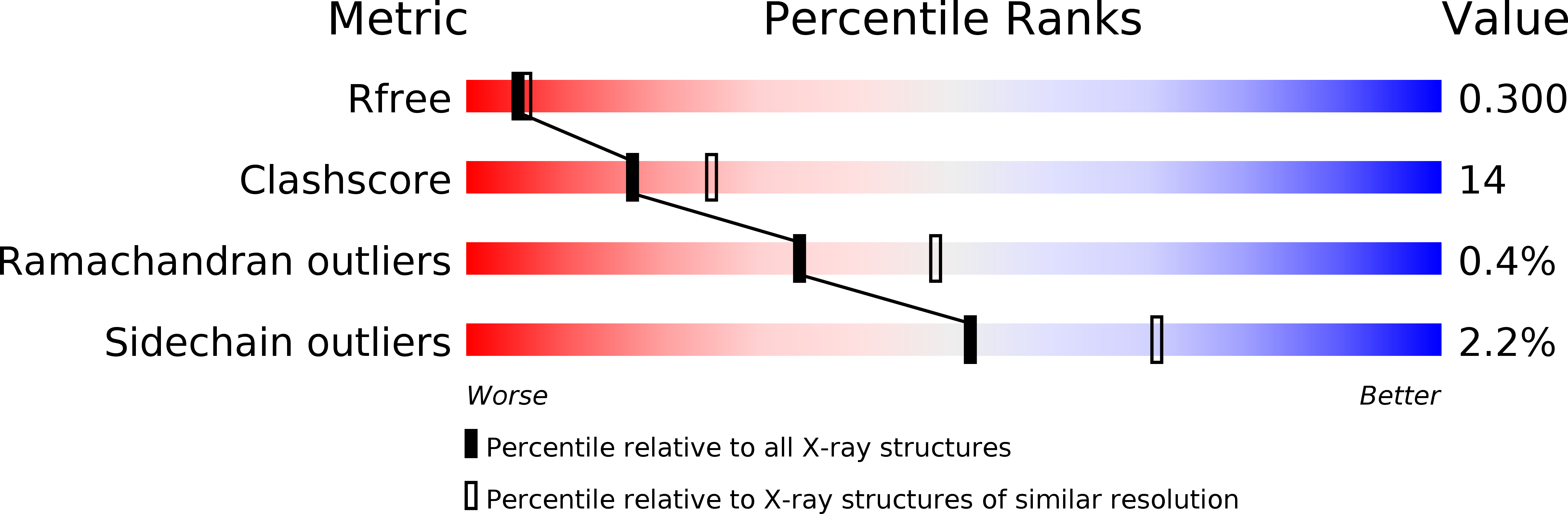
Resolution:
2.39 Å
R-Value Free:
0.23
R-Value Work:
0.18
R-Value Observed:
0.19
Space Group:
P 32