2XZR image
Deposition Date 2010-11-28
Release Date 2011-07-20
Last Version Date 2023-12-20
Entry Detail
PDB ID:
2XZR
Keywords:
Title:
Escherichia coli Immunoglobulin-binding protein EibD 391-438 FUSED TO GCN4 ADAPTORS
Biological Source:
Source Organism:
Host Organism:
Method Details:
Experimental Method:
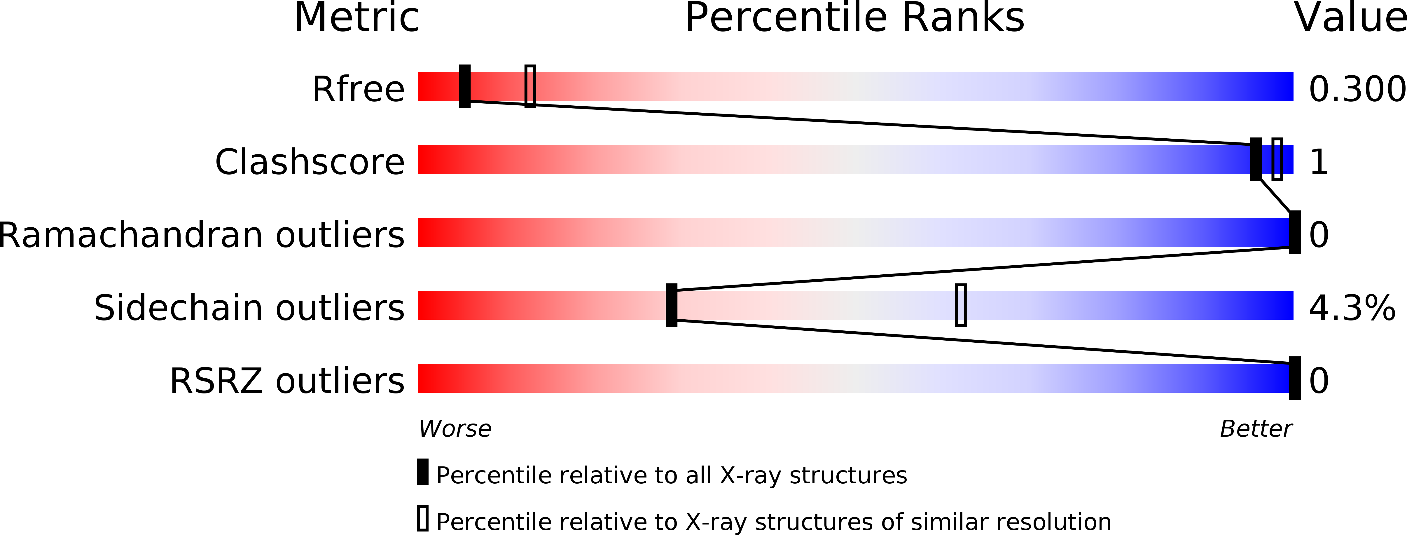
Resolution:
2.80 Å
R-Value Free:
0.30
R-Value Work:
0.25
R-Value Observed:
0.26
Space Group:
P 3 2 1
Macromolecular Entities
Polymer Type:polypeptide(L)
Molecule:IMMUNOGLOBULIN-BINDING PROTEIN EIBD
Gene (Uniprot):eibD
Chain IDs:A
Chain Length:114
Number of Molecules:1
Biological Source:ENTEROBACTERIA PHAGE P-EIBD
Ligand Molecules
Primary Citation
The Structure of E. Coli Igg-Binding Protein D Suggests a General Model for Bending and Binding in Trimeric Autotransporter Adhesins.
Structure 19 1021 ? (2011)
PMID: 21742268 DOI: 10.1016/J.STR.2011.03.021

Abstact

The Escherichia coli Ig-binding (Eib) proteins are trimeric autotransporter adhesins (TAAs) and receptors for IgG Fc. We present the structure of a large fragment of the passenger domain of EibD, the first TAA structure to have both a YadA-like head domain and the entire coiled-coil stalk. The stalk begins as a right-handed superhelix, but switches handedness halfway down. An unexpected β-minidomain joins the two and inserts a ∼120° rotation such that there is no net twist between the beginning and end of the stalk. This may be important in folding and autotransport. The surprisingly large cavities we found in EibD and other TAAs may explain how TAAs bend to bind their ligands. We identified how IgA and IgG bind and modeled the EibD-IgG Fc complex. We further show that EibD promotes autoagglutination and biofilm formation and forms a fibrillar layer covering the cell surface making zipper-like contacts between cells.

Legend

Protein

Chemical

Disease

Primary Citation of related structures