2XZE image
Deposition Date 2010-11-25
Release Date 2011-08-24
Last Version Date 2024-05-08
Entry Detail
PDB ID:
2XZE
Title:
Structural basis for AMSH-ESCRT-III CHMP3 interaction
Biological Source:
Source Organism:
HOMO SAPIENS (Taxon ID: 9606)
Host Organism:
Method Details:
Experimental Method:
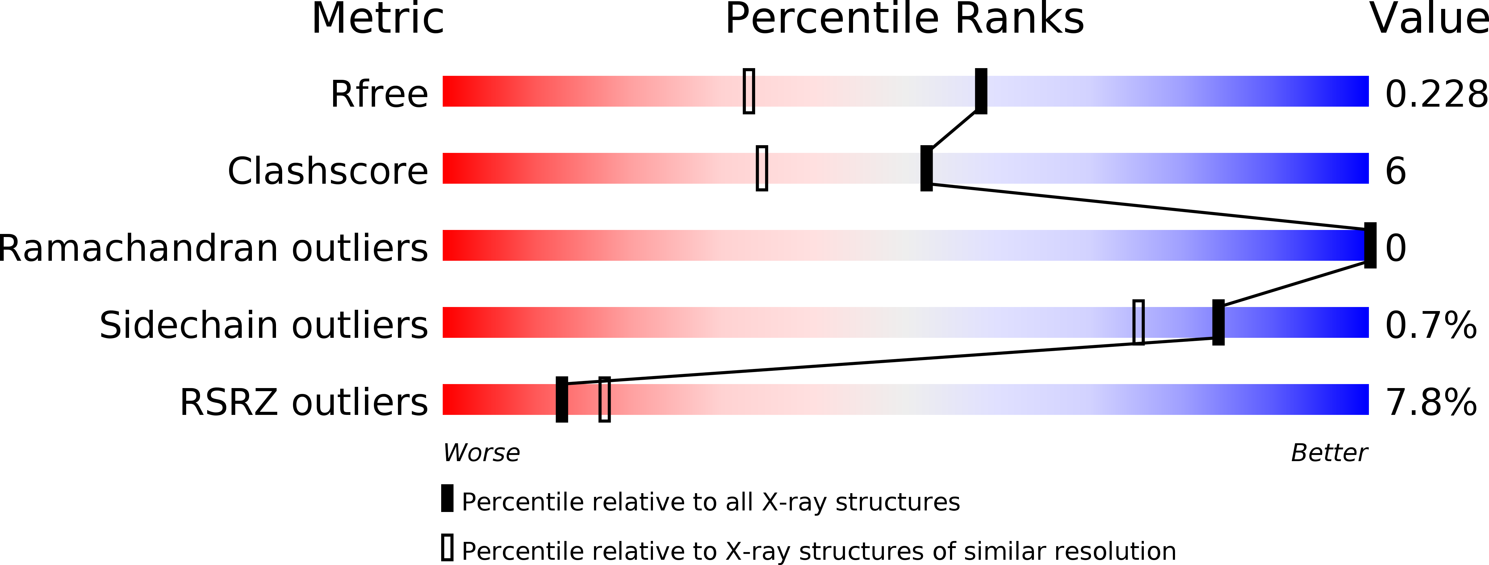
Resolution:
1.75 Å
R-Value Free:
0.22
R-Value Work:
0.19
R-Value Observed:
0.19
Space Group:
P 41
Macromolecular Entities
Structures with similar UniProt ID
Protein Blast
Polymer Type:polypeptide(L)
Molecule:STAM-BINDING PROTEIN
Gene (Uniprot):STAMBP
Chain IDs:A, B
Chain Length:146
Number of Molecules:2
Biological Source:HOMO SAPIENS
Structures with similar UniProt ID
Protein Blast
Polymer Type:polypeptide(L)
Molecule:CHARGED MULTIVESICULAR BODY PROTEIN 3
Gene (Uniprot):CHMP3
Chain IDs:C (auth: Q), D (auth: R)
Chain Length:40
Number of Molecules:2
Biological Source:HOMO SAPIENS
Primary Citation

Abstact

Endosomal sorting complexes required for transport (ESCRT) recognize ubiquitinated cargo and catalyze diverse budding processes including multivesicular body biogenesis, enveloped virus egress, and cytokinesis. We present the crystal structure of an N-terminal fragment of the deubiquitinating enzyme AMSH (AMSHΔC) in complex with the C-terminal region of ESCRT-III CHMP3 (CHMP3ΔN). AMSHΔC folds into an elongated 90 Å long helical assembly that includes an unusual MIT domain. CHMP3ΔN is unstructured in solution and helical in complex with AMSHΔC, revealing a novel MIT domain interacting motif (MIM) that does not overlap with the CHMP1-AMSH binding site. ITC and SPR measurements demonstrate an unusual high-affinity MIM-MIT interaction. Structural analysis suggests a regulatory role for the N-terminal helical segment of AMSHΔC and its destabilization leads to a loss of function during HIV-1 budding. Our results indicate a tight coupling of ESCRT-III CHMP3 and AMSH functions and provide insight into the regulation of ESCRT-III.

Legend

Protein

Chemical

Disease

Primary Citation of related structures