2XUL image
Deposition Date 2010-10-19
Release Date 2010-11-03
Last Version Date 2024-05-08
Entry Detail
PDB ID:
2XUL
Title:
Structure of PII from Synechococcus elongatus in complex with 2- oxoglutarate at high 2-OG concentrations
Biological Source:
Source Organism:
Host Organism:
Method Details:
Experimental Method:
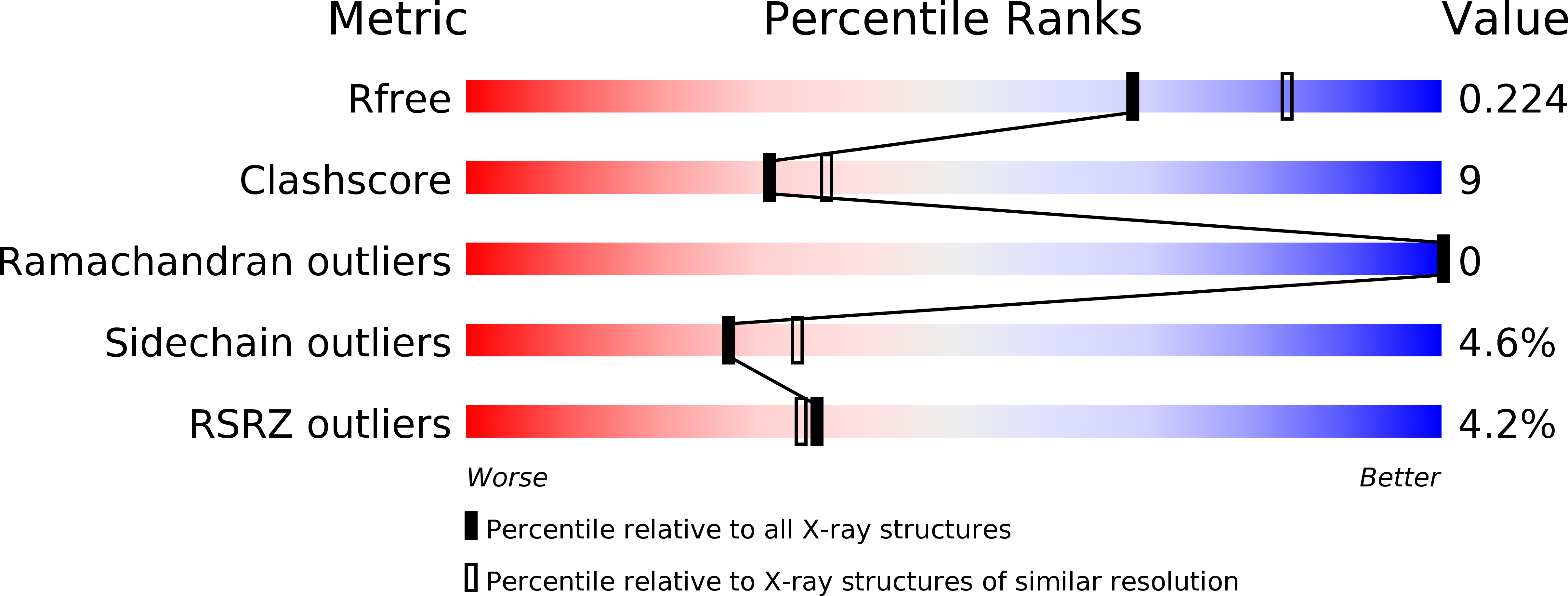
Resolution:
2.20 Å
R-Value Free:
0.22
R-Value Work:
0.17
R-Value Observed:
0.18
Space Group:
P 21 21 21
Macromolecular Entities
Structures with similar UniProt ID
Protein Blast
Polymer Type:polypeptide(L)
Molecule:NITROGEN REGULATORY PROTEIN P-II
Gene (Uniprot):glnB
Chain IDs:A, B, C, D, E, F
Chain Length:115
Number of Molecules:6
Biological Source:SYNECHOCOCCUS ELONGATUS
Primary Citation
Mechanism of 2-Oxoglutarate Signaling by the Synechococcus Elongatus Pii Signal Transduction Protein
Proc.Natl.Acad.Sci.USA 107 19760 ? (2010)
PMID: 21041661 DOI: 10.1073/PNAS.1007653107

Abstact

P(II) proteins control key processes of nitrogen metabolism in bacteria, archaea, and plants in response to the central metabolites ATP, ADP, and 2-oxoglutarate (2-OG), signaling cellular energy and carbon and nitrogen abundance. This metabolic information is integrated by P(II) and transmitted to regulatory targets (key enzymes, transporters, and transcription factors), modulating their activity. In oxygenic phototrophs, the controlling enzyme of arginine synthesis, N-acetyl-glutamate kinase (NAGK), is a major P(II) target, whose activity responds to 2-OG via P(II). Here we show structures of the Synechococcus elongatus P(II) protein in complex with ATP, Mg(2+), and 2-OG, which clarify how 2-OG affects P(II)-NAGK interaction. P(II) trimers with all three sites fully occupied were obtained as well as structures with one or two 2-OG molecules per P(II) trimer. These structures identify the site of 2-OG located in the vicinity between the subunit clefts and the base of the T loop. The 2-OG is bound to a Mg(2+) ion, which is coordinated by three phosphates of ATP, and by ionic interactions with the highly conserved residues K58 and Q39 together with B- and T-loop backbone interactions. These interactions impose a unique T-loop conformation that affects the interactions with the P(II) target. Structures of P(II) trimers with one or two bound 2-OG molecules reveal the basis for anticooperative 2-OG binding and shed light on the intersubunit signaling mechanism by which P(II) senses effectors in a wide range of concentrations.

Legend

Protein

Chemical

Disease

Primary Citation of related structures