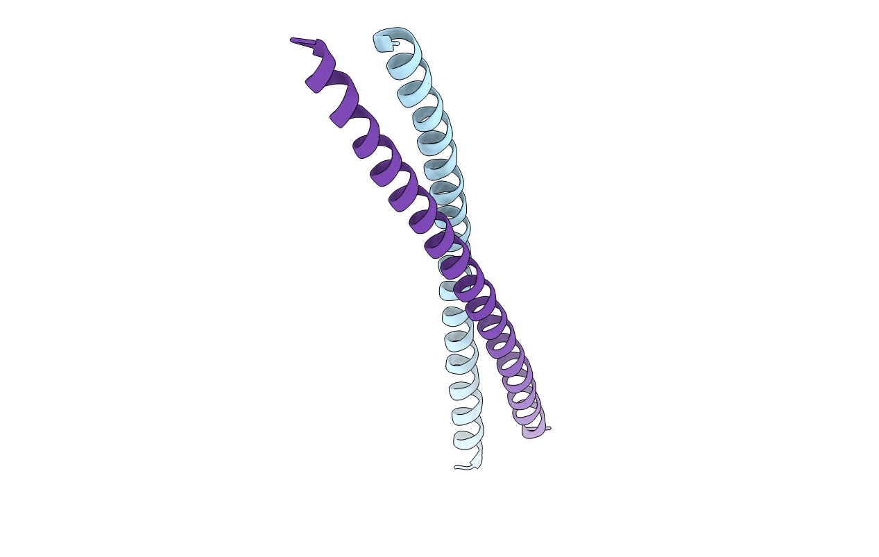
2XU6 image
Deposition Date 2010-10-14
Release Date 2010-10-27
Last Version Date 2024-11-06
Entry Detail
PDB ID:
2XU6
Keywords:
Title:
MDV1 coiled coil domain
Biological Source:
Source Organism:
Host Organism:
Method Details:
Experimental Method:
Resolution:
2.70 Å
R-Value Free:
0.31
R-Value Work:
0.27
R-Value Observed:
0.27
Space Group:
P 41 21 2
Macromolecular Entities
Polymer Type:polypeptide(L)
Molecule:MDV1 COILED COIL
Gene (Uniprot):MDV1
Mutations:YES
Chain IDs:A, B
Chain Length:72
Number of Molecules:2
Biological Source:SACCHAROMYCES CEREVISIAE
Modified Residue
Compound ID Chain ID Parent Comp ID Details 2D Image
MSE A MET SELENOMETHIONINE
Primary Citation
Molecular Architecture of a Dynamin Adaptor: Implications for Assembly of Mitochondrial Fission Complexes
J.Cell Biol. 191 1127 ? (2010)
PMID: 21149566 DOI: 10.1083/JCB.201005046

Abstact

Recruitment and assembly of some dynamin-related guanosine triphosphatases depends on adaptor proteins restricted to distinct cellular membranes. The yeast Mdv1 adaptor localizes to mitochondria by binding to the membrane protein Fis1. Subsequent Mdv1 binding to the mitochondrial dynamin Dnm1 stimulates Dnm1 assembly into spirals, which encircle and divide the mitochondrial compartment. In this study, we report that dimeric Mdv1 is joined at its center by a 92-Å antiparallel coiled coil (CC). Modeling of the Fis1-Mdv1 complex using available crystal structures suggests that the Mdv1 CC lies parallel to the bilayer with N termini at opposite ends bound to Fis1 and C-terminal β-propeller domains (Dnm1-binding sites) extending into the cytoplasm. A CC length of appropriate length and sequence is necessary for optimal Mdv1 interaction with Fis1 and Dnm1 and is important for proper Dnm1 assembly before membrane scission. Our results provide a framework for understanding how adaptors act as scaffolds to orient and stabilize the assembly of dynamins on membranes.

Legend

Protein

Chemical

Disease

Primary Citation of related structures