Deposition Date
2010-10-01
Release Date
2011-10-05
Last Version Date
2024-11-20
Entry Detail
PDB ID:
2XSZ
Keywords:
Title:
The dodecameric human RuvBL1:RuvBL2 complex with truncated domains II
Biological Source:
Source Organism(s):
HOMO SAPIENS (Taxon ID: 9606)
Expression System(s):
Method Details:
Experimental Method:
Resolution:
3.00 Å
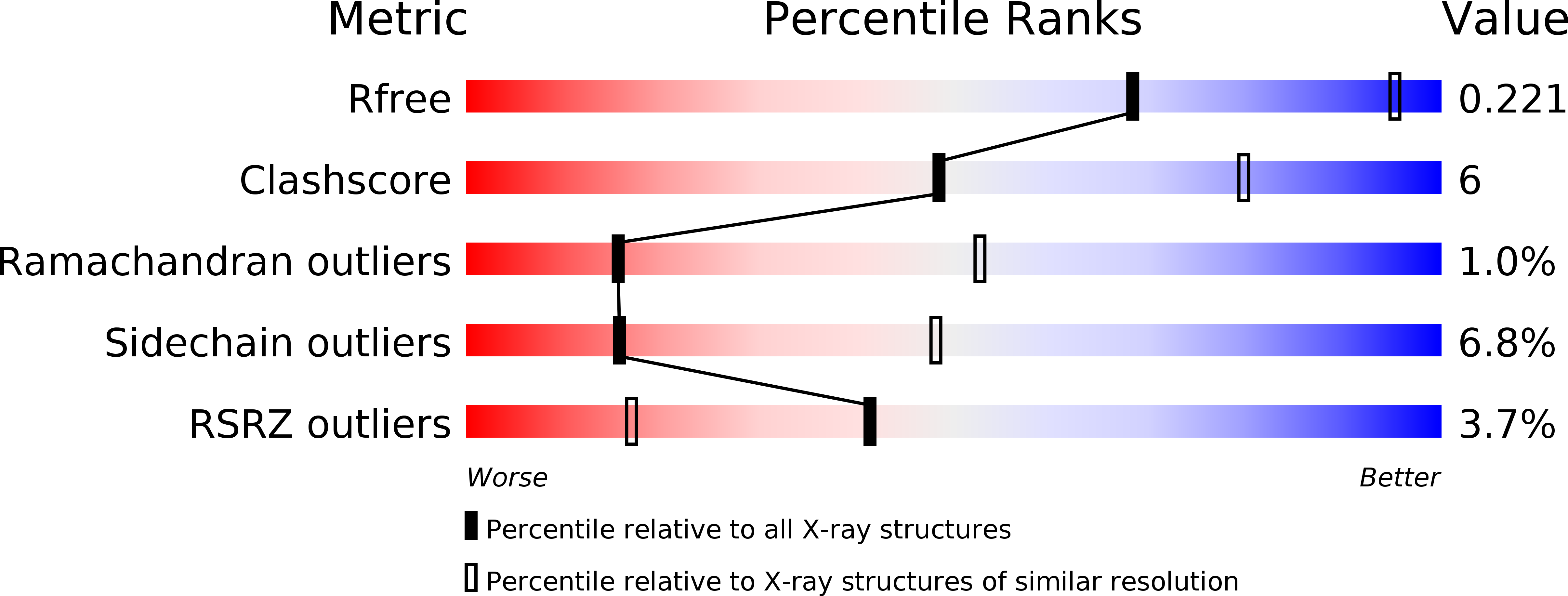
R-Value Free:
0.20
R-Value Work:
0.17
R-Value Observed:
0.17
Space Group:
C 2 2 21