2XSJ image
Deposition Date 2010-09-29
Release Date 2011-08-24
Last Version Date 2024-11-13
Entry Detail
PDB ID:
2XSJ
Keywords:
Title:
Structure of desulforubidin from Desulfomicrobium norvegicum
Biological Source:
Source Organism:
Method Details:
Experimental Method:
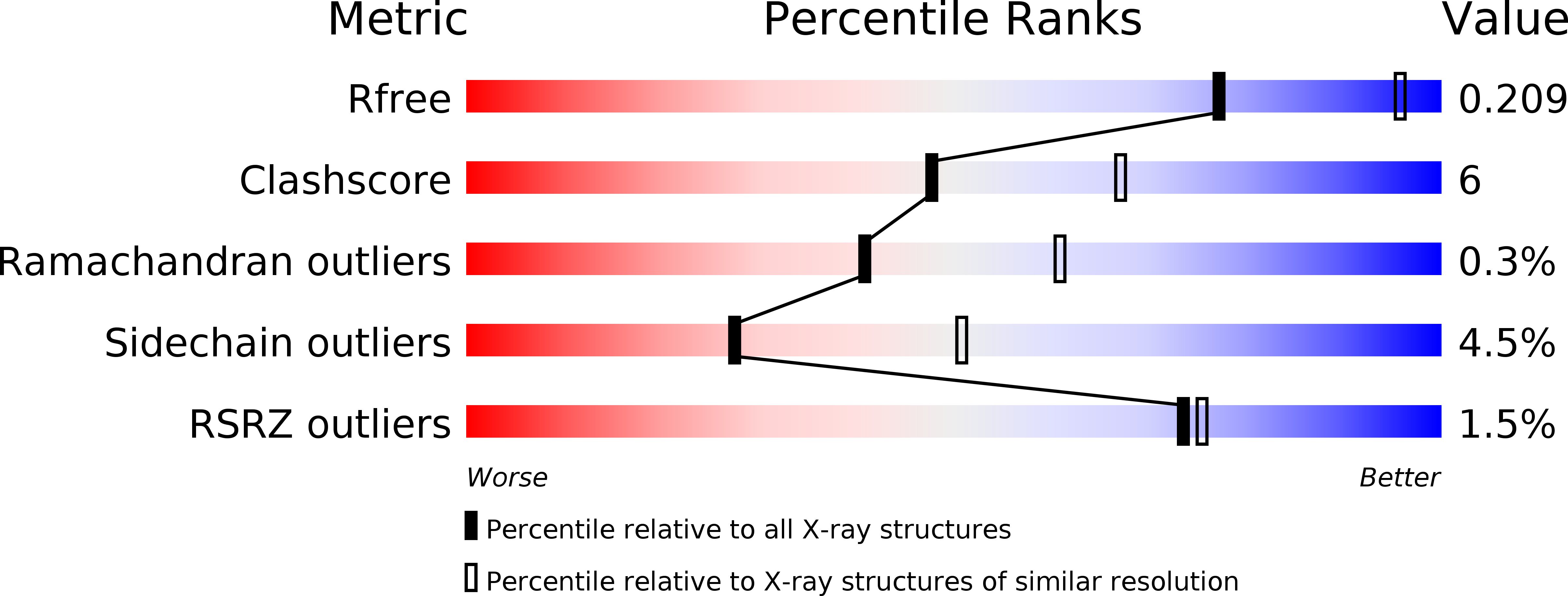
Resolution:
2.50 Å
R-Value Free:
0.20
R-Value Work:
0.15
R-Value Observed:
0.15
Space Group:
P 21 21 21
Macromolecular Entities
Polymer Type:polypeptide(L)
Molecule:SULFITE REDUCTASE ALPHA SUBUNIT
Gene (Uniprot):dsrA
Chain IDs:A, D
Chain Length:437
Number of Molecules:2
Biological Source:DESULFOMICROBIUM NORVEGICUM
Polymer Type:polypeptide(L)
Molecule:SULFITE REDUCTASE BETA SUBUNIT
Gene (Uniprot):dsrB
Chain IDs:B, E
Chain Length:386
Number of Molecules:2
Biological Source:DESULFOMICROBIUM NORVEGICUM
Polymer Type:polypeptide(L)
Molecule:SULFUR RELAY PROTEIN, TUSE/DSRC/DSVC FAMILY
Chain IDs:C, F
Chain Length:105
Number of Molecules:2
Biological Source:DESULFOMICROBIUM NORVEGICUM
Primary Citation
Structural Insights Into Dissimilatory Sulfite Reductases: Structure of Desulforubidin from Desulfomicrobium Norvegicum
Front.Microbiol. 2 71 ? (2011)
PMID: 21833321 DOI: 10.3389/FMICB.2011.00071

Abstact

Dissimilatory sulfite reductases (dSiRs) are crucial enzymes in bacterial sulfur-based energy metabolism, which are likely to have been present in some of the earliest life forms on Earth. Several classes of dSiRs have been proposed on the basis of different biochemical and spectroscopic properties, but it is not clear whether this corresponds to actual physiological or structural differences. Here, we describe the first structure of a dSiR from the desulforubidin class isolated from Desulfomicrobium norvegicum. The desulforubidin (Drub) structure is assembled as α(2)β(2)γ(2), in which two DsrC proteins are bound to the core [DsrA](2)[DsrB](2) unit, as reported for the desulfoviridin (Dvir) structure from Desulfovibrio vulgaris. Unlike Dvir, four sirohemes and eight [4Fe-4S] clusters are present in Drub. However, the structure indicates that only two of the Drub coupled siroheme-[4Fe-4S] cofactors are catalytically active. Mass spectrometry studies of purified Drub and Dvir show that both proteins present different oligomeric complex forms that bind two, one, or no DsrC proteins, providing an explanation for conflicting spectroscopic and biochemical results in the literature, and further indicating that DsrC is not a subunit of dSiR, but rather a protein with which it interacts.

Legend

Protein

Chemical

Disease

Primary Citation of related structures