Deposition Date
2010-09-24
Release Date
2010-11-03
Last Version Date
2023-12-20
Entry Detail
PDB ID:
2XS8
Keywords:
Title:
Crystal Structure of ALIX in complex with the SIVagmTan-1 AYDPARKLL Late Domain
Biological Source:
Source Organism(s):
HOMO SAPIENS (Taxon ID: 9606)
SIMIAN IMMUNODEFICIENCY VIRUS (Taxon ID: 11723)
SIMIAN IMMUNODEFICIENCY VIRUS (Taxon ID: 11723)
Expression System(s):
Method Details:
Experimental Method:
Resolution:
2.50 Å
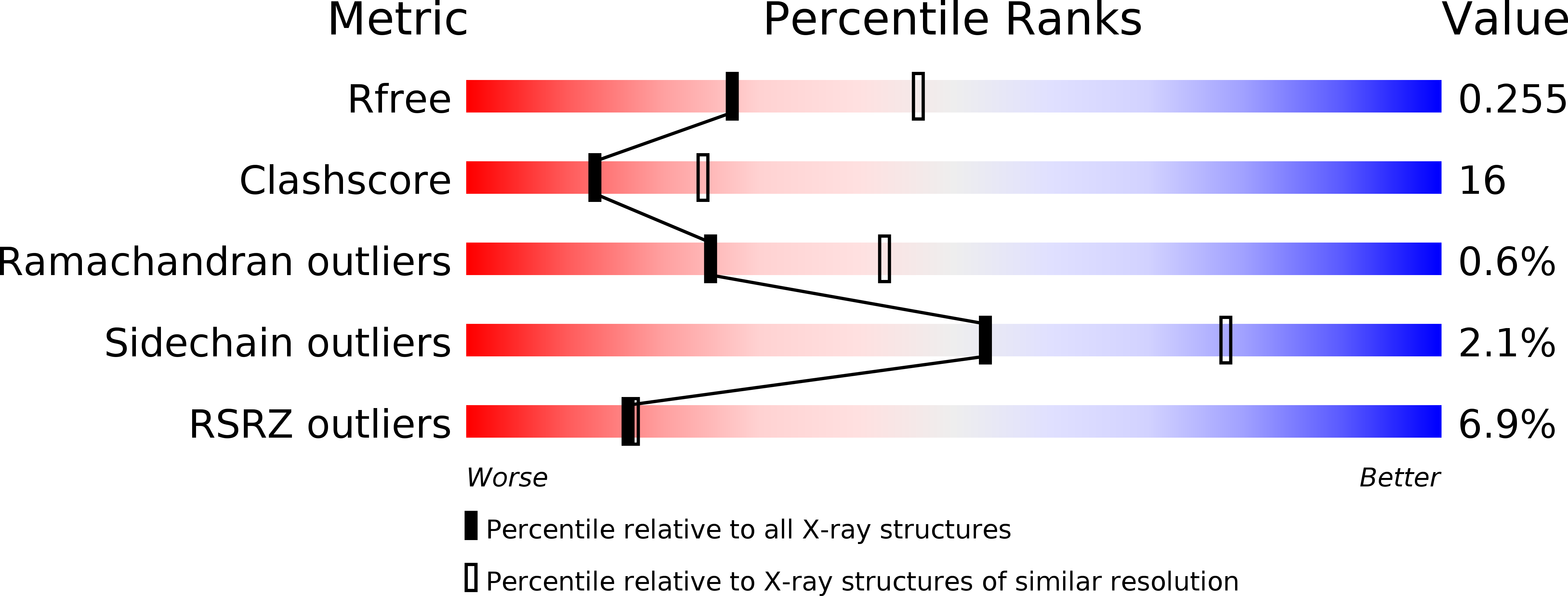
R-Value Free:
0.26
R-Value Work:
0.20
R-Value Observed:
0.20
Space Group:
C 1 2 1