2XRZ image
Entry Detail
PDB ID:
2XRZ
Keywords:
Title:
X-ray structure of archaeal class II CPD photolyase from Methanosarcina mazei in complex with intact CPD-lesion
Biological Source:
Host Organism:
PDB Version:
Deposition Date:
2010-09-24
Release Date:
2011-09-14
Method Details:
Experimental Method:
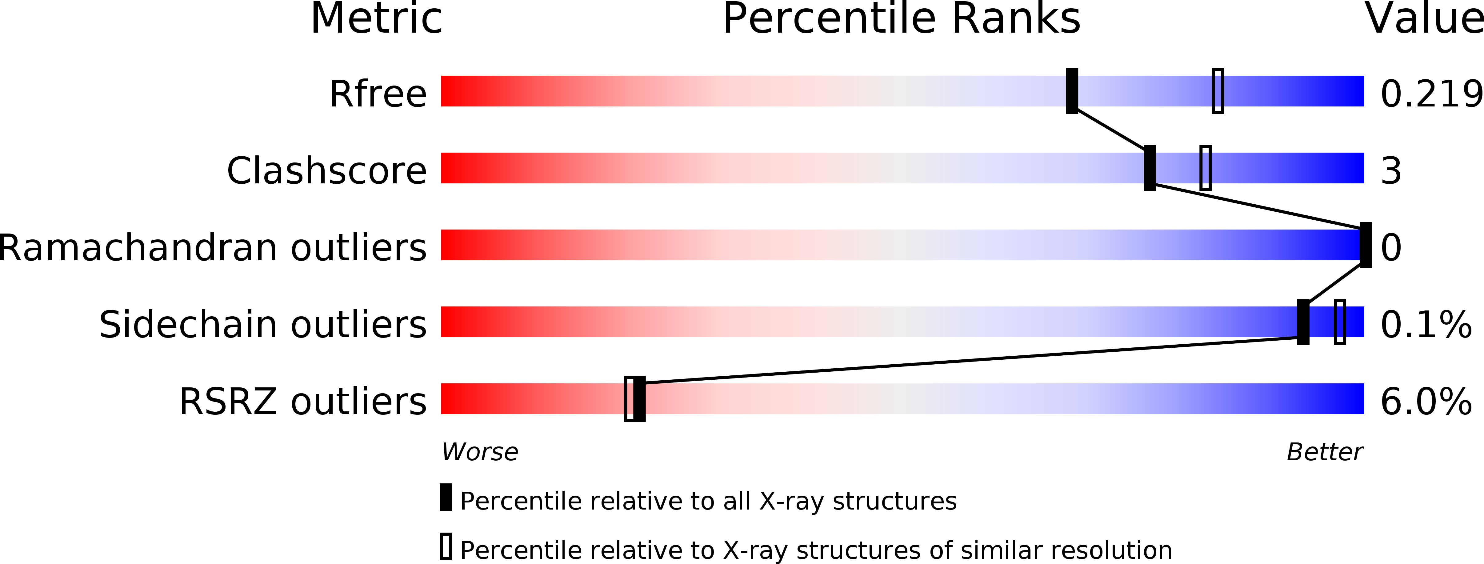
Resolution:
2.20 Å
R-Value Free:
0.20
R-Value Work:
0.17
R-Value Observed:
0.17
Space Group:
P 21 21 21
Macromolecular Entities
Polymer Type:polypeptide(L)
Description:DEOXYRIBODIPYRIMIDINE PHOTOLYASE
Mutations:YES
Chain IDs:A, B
Chain Length:482
Number of Molecules:2
Biological Source:METHANOSARCINA MAZEI
Polymer Type:polydeoxyribonucleotide
Description:CPD-COMPRISING OLIGONUCLEOTIDE
Chain IDs:C, E
Chain Length:14
Number of Molecules:2
Biological Source:SYNTHETIC CONSTRUCT
Polymer Type:polydeoxyribonucleotide
Description:COUNTERSTRAND-OLIGONUCLEOTIDE
Chain IDs:D, F
Chain Length:14
Number of Molecules:2
Biological Source:SYNTHETIC CONSTRUCT
Primary Citation
Crystal Structures of an Archaeal Class II DNA Photolyase and its Complex with Uv-Damaged Duplex DNA.
Embo J. 30 4437 ? (2011)
PMID: 21892138 DOI: 10.1038/EMBOJ.2011.313

Abstact

Class II photolyases ubiquitously occur in plants, animals, prokaryotes and some viruses. Like the distantly related microbial class I photolyases, these enzymes repair UV-induced cyclobutane pyrimidine dimer (CPD) lesions within duplex DNA using blue/near-UV light. Methanosarcina mazei Mm0852 is a class II photolyase of the archaeal order of Methanosarcinales, and is closely related to plant and metazoan counterparts. Mm0852 catalyses light-driven DNA repair and photoreduction, but in contrast to class I enzymes lacks a high degree of binding discrimination between UV-damaged and intact duplex DNA. We solved crystal structures of Mm0852, the first one for a class II photolyase, alone and in complex with CPD lesion-containing duplex DNA. The lesion-binding mode differs from other photolyases by a larger DNA-binding site, and an unrepaired CPD lesion is found flipped into the active site and recognized by a cluster of five water molecules next to the bound 3'-thymine base. Different from other members of the photolyase-cryptochrome family, class II photolyases appear to utilize an unusual, conserved tryptophane dyad as electron transfer pathway to the catalytic FAD cofactor.

Legend

Protein

Chemical

Disease

Primary Citation of related structures