Deposition Date
2010-08-31
Release Date
2010-12-29
Last Version Date
2023-12-20
Entry Detail
PDB ID:
2XPY
Keywords:
Title:
Structure of Native Leukotriene A4 Hydrolase from Saccharomyces cerevisiae
Biological Source:
Source Organism(s):
SACCHAROMYCES CEREVISIAE (Taxon ID: 4932)
Expression System(s):
Method Details:
Experimental Method:
Resolution:
2.73 Å
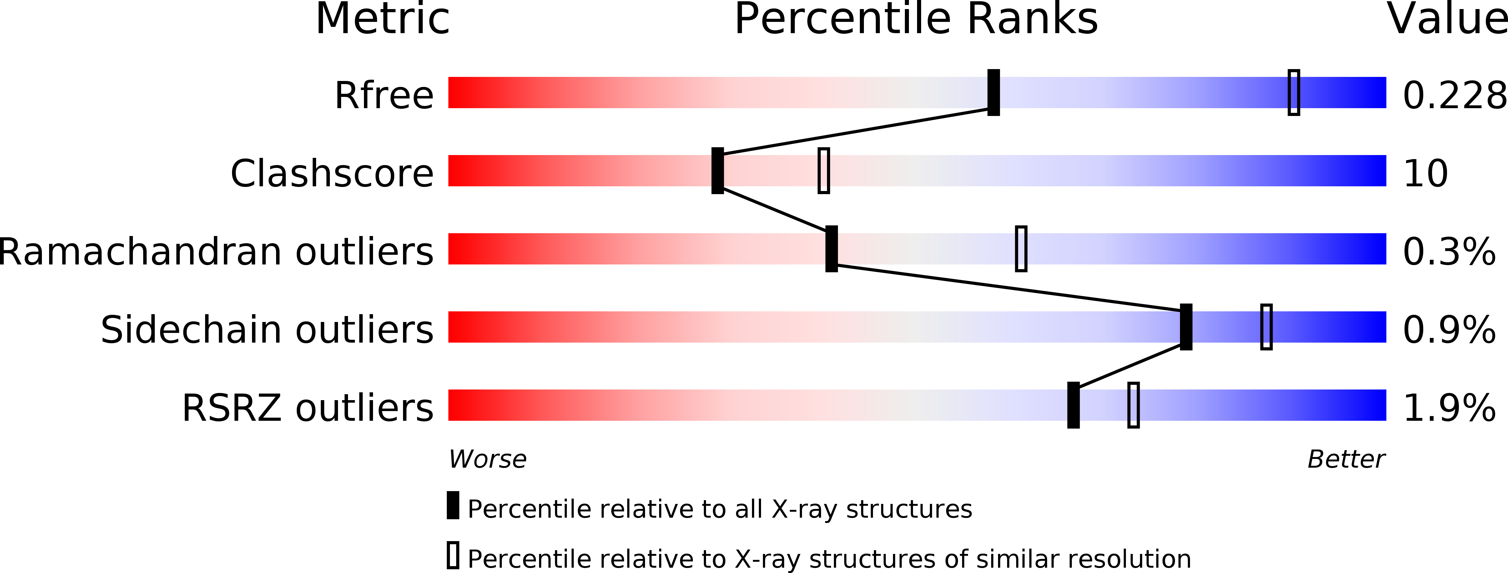
R-Value Free:
0.24
R-Value Work:
0.17
R-Value Observed:
0.17
Space Group:
P 32 2 1