2XO4 image
Deposition Date 2010-08-09
Release Date 2010-08-18
Last Version Date 2024-11-20
Entry Detail
PDB ID:
2XO4
Keywords:
Title:
RIBONUCLEOTIDE REDUCTASE Y730NH2Y MODIFIED R1 SUBUNIT OF E. COLI
Biological Source:
Source Organism:
Host Organism:
Method Details:
Experimental Method:
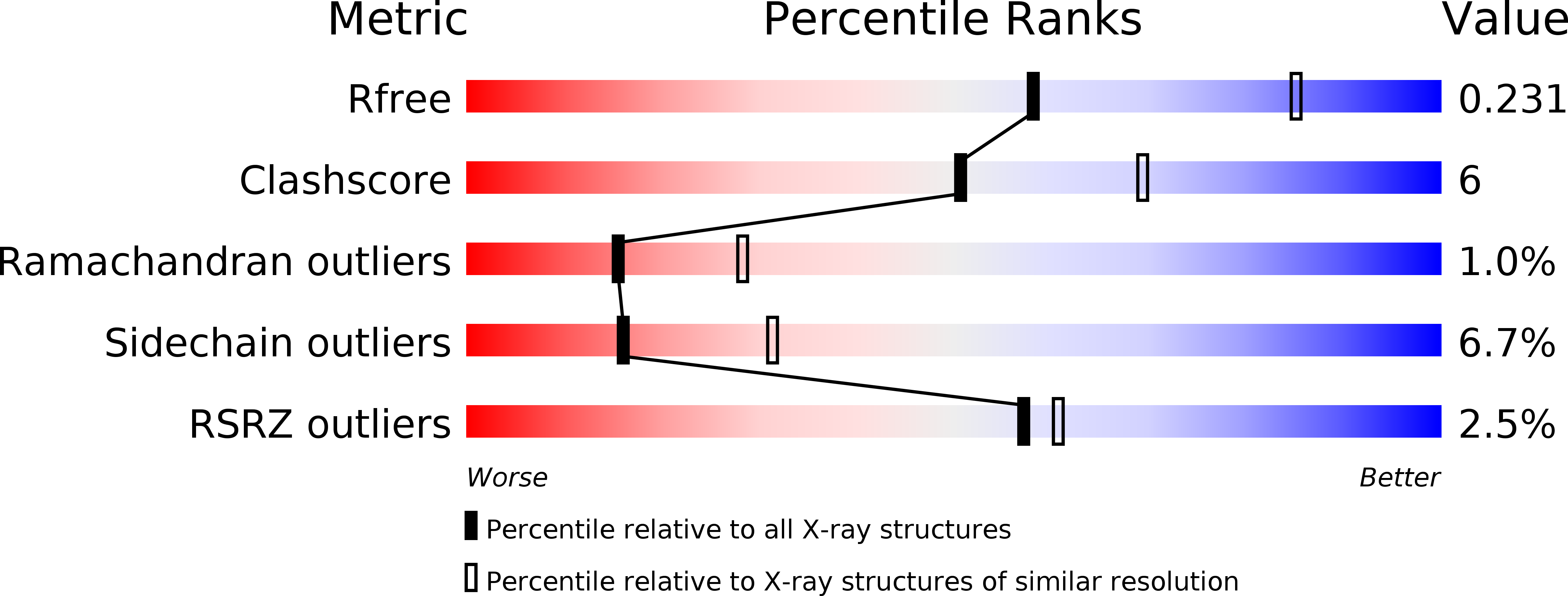
Resolution:
2.50 Å
R-Value Free:
0.23
R-Value Work:
0.18
R-Value Observed:
0.19
Space Group:
H 3 2
Macromolecular Entities
Structures with similar UniProt ID
Protein Blast
Polymer Type:polypeptide(L)
Molecule:RIBONUCLEOSIDE-DIPHOSPHATE REDUCTASE 1 SUBUNIT ALPHA
Gene (Uniprot):nrdA
Chain IDs:A, B, C
Chain Length:761
Number of Molecules:3
Biological Source:ESCHERICHIA COLI
Structures with similar UniProt ID
Protein Blast
Polymer Type:polypeptide(L)
Molecule:RIBONUCLEOSIDE-DIPHOSPHATE REDUCTASE 1 SUBUNIT BETA
Gene (Uniprot):nrdB
Chain IDs:D, E, F, G (auth: P)
Chain Length:20
Number of Molecules:4
Biological Source:ESCHERICHIA COLI
Modified Residue
Compound ID Chain ID Parent Comp ID Details 2D Image
TY2 A TYR 3-AMINO-L-TYROSINE
Primary Citation
Kinetics of Radical Intermediate Formation and Deoxynucleotide Production in 3-Aminotyrosine- Substituted Escherichia Coli Ribonucleotide Reductases.
J.Am.Chem.Soc. 133 9430 ? (2011)
PMID: 21612216 DOI: 10.1021/JA201640N

Abstact

Escherichia coli ribonucleotide reductase is an α2β2 complex and catalyzes the conversion of nucleoside 5'-diphosphates (NDPs) to 2'-deoxynucleotides (dNDPs). The reaction is initiated by the transient oxidation of an active-site cysteine (C(439)) in α2 by a stable diferric tyrosyl radical (Y(122)•) cofactor in β2. This oxidation occurs by a mechanism of long-range proton-coupled electron transfer (PCET) over 35 Å through a specific pathway of residues: Y(122)•→ W(48)→ Y(356) in β2 to Y(731)→ Y(730)→ C(439) in α2. To study the details of this process, 3-aminotyrosine (NH(2)Y) has been site-specifically incorporated in place of Y(356) of β. The resulting protein, Y(356)NH(2)Y-β2, and the previously generated proteins Y(731)NH(2)Y-α2 and Y(730)NH(2)Y-α2 (NH(2)Y-RNRs) are shown to catalyze dNDP production in the presence of the second subunit, substrate (S), and allosteric effector (E) with turnover numbers of 0.2-0.7 s(-1). Evidence acquired by three different methods indicates that the catalytic activity is inherent to NH(2)Y-RNRs and not the result of copurifying wt enzyme. The kinetics of formation of 3-aminotyrosyl radical (NH(2)Y•) at position 356, 731, and 730 have been measured with all S/E pairs. In all cases, NH(2)Y• formation is biphasic (k(fast) of 9-46 s(-1) and k(slow) of 1.5-5.0 s(-1)) and kinetically competent to be an intermediate in nucleotide reduction. The slow phase is proposed to report on the conformational gating of NH(2)Y• formation, while the k(cat) of ~0.5 s(-1) is proposed to be associated with rate-limiting oxidation by NH(2)Y• of the subsequent amino acid on the pathway during forward PCET. The X-ray crystal structures of Y(730)NH(2)Y-α2 and Y(731)NH(2)Y-α2 have been solved and indicate minimal structural changes relative to wt-α2. From the data, a kinetic model for PCET along the radical propagation pathway is proposed.

Legend

Protein

Chemical

Disease