Deposition Date
2010-07-30
Release Date
2011-08-10
Last Version Date
2024-05-08
Entry Detail
PDB ID:
2XN0
Keywords:
Title:
Structure of alpha-galactosidase from Lactobacillus acidophilus NCFM, PtCl4 derivative
Biological Source:
Source Organism(s):
LACTOBACILLUS ACIDOPHILUS NCFM (Taxon ID: 272621)
Expression System(s):
Method Details:
Experimental Method:
Resolution:
2.50 Å
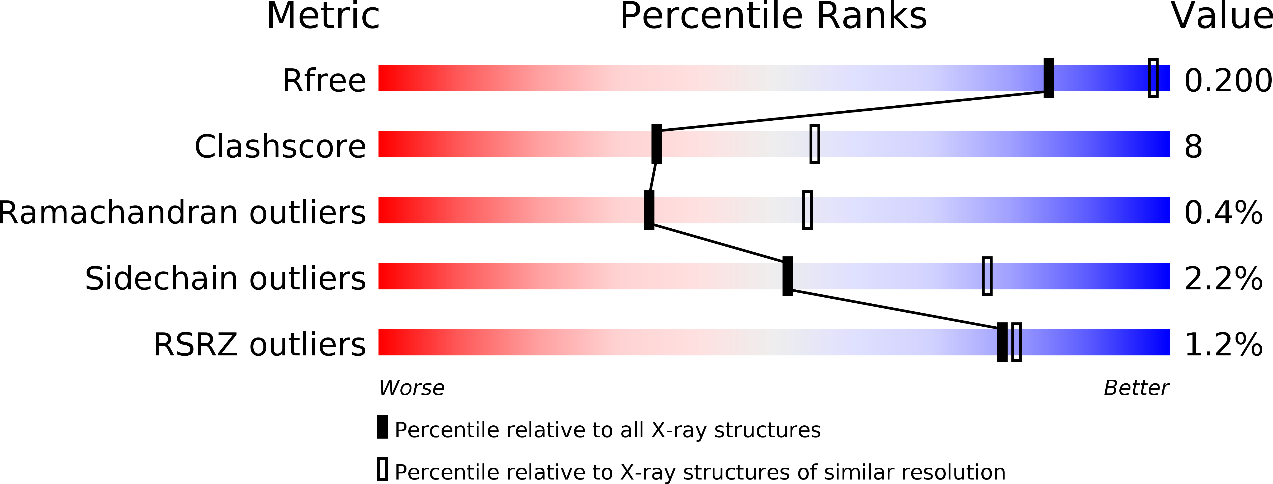
R-Value Free:
0.20
R-Value Work:
0.14
R-Value Observed:
0.15
Space Group:
P 21 21 2