2XLC image
Entry Detail
PDB ID:
2XLC
Keywords:
Title:
Acetyl xylan esterase from Bacillus pumilus CECT5072 bound to paraoxon
Biological Source:
Source Organism:
Host Organism:
PDB Version:
Deposition Date:
2010-07-20
Release Date:
2011-05-25
Method Details:
Experimental Method:
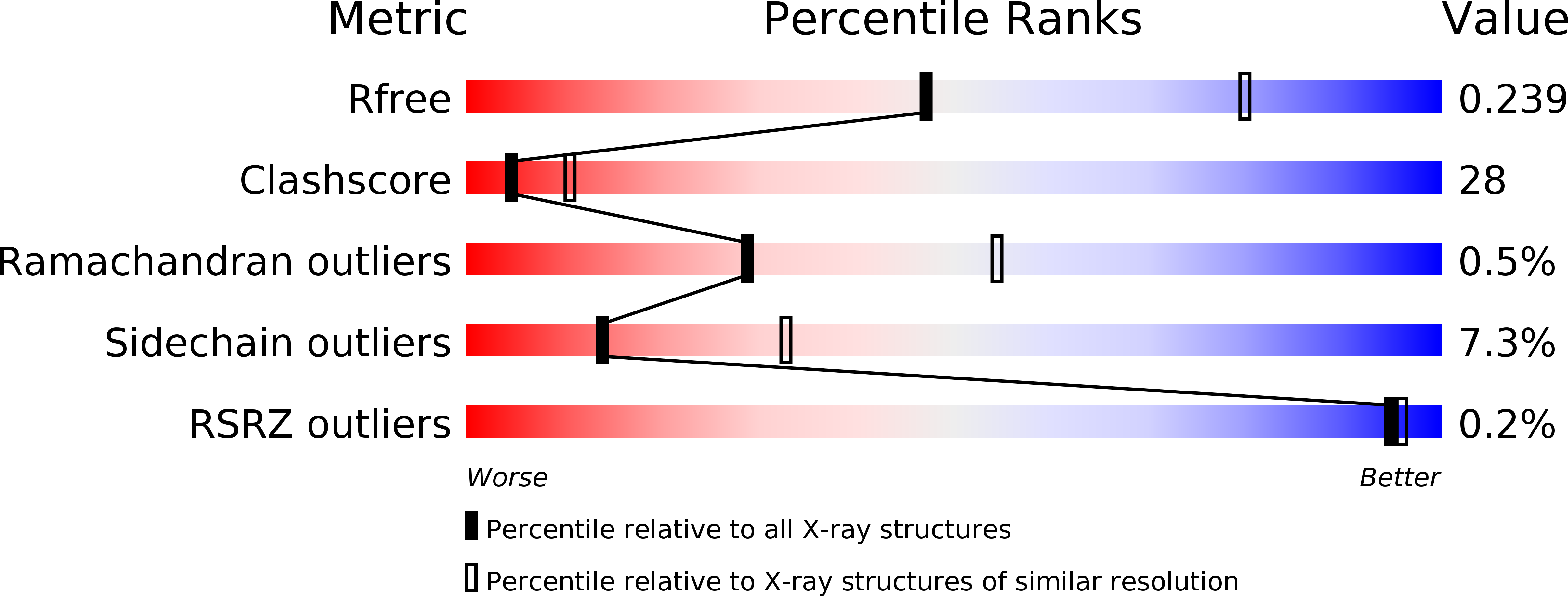
Resolution:
2.70 Å
R-Value Free:
0.23
R-Value Work:
0.21
R-Value Observed:
0.21
Space Group:
P 1 21 1
Macromolecular Entities
Polymer Type:polypeptide(L)
Description:ACETYL XYLAN ESTERASE
Chain IDs:A, B, C, D, E, F
Chain Length:320
Number of Molecules:6
Biological Source:BACILLUS PUMILUS
Modified Residue
Compound ID Chain ID Parent Comp ID Details 2D Image
MSE A MET SELENOMETHIONINE
Ligand Molecules
Primary Citation
The Crystal Structure of the Cephalosporin Deacetylating Enzyme Acetyl Xylan Esterase Bound to Paraoxon Explains the Low Sensitivity of This Serine Hydrolase to Organophosphate Inactivation.
Biochem.J. 436 321 ? (2011)
PMID: 21382014 DOI: 10.1042/BJ20101859

Abstact

Organophosphorus insecticides and nerve agents irreversibly inhibit serine hydrolase superfamily enzymes. One enzyme of this superfamily, the industrially important (for β-lactam antibiotic synthesis) AXE/CAH (acetyl xylan esterase/cephalosporin acetyl hydrolase) from the biotechnologically valuable organism Bacillus pumilus, exhibits low sensitivity to the organophosphate paraoxon (diethyl-p-nitrophenyl phosphate, also called paraoxon-ethyl), reflected in a high K(i) for it (~5 mM) and in a slow formation (t(½)~1 min) of the covalent adduct of the enzyme and for DEP (E-DEP, enzyme-diethyl phosphate, i.e. enzyme-paraoxon). The crystal structure of the E-DEP complex determined at 2.7 Å resolution (1 Å=0.1 nm) reveals strain in the active Ser¹⁸¹-bound organophosphate as a likely cause for the limited paraoxon sensitivity. The strain results from active-site-size limitation imposed by bulky conserved aromatic residues that may exclude as substrates esters having acyl groups larger than acetate. Interestingly, in the doughnut-like homohexamer of the enzyme, the six active sites are confined within a central chamber formed between two 60°-staggered trimers. The exclusive access to this chamber through a hole around the three-fold axis possibly limits the size of the xylan natural substrates. The enzyme provides a rigid scaffold for catalysis, as reflected in the lack of movement associated with paraoxon adduct formation, as revealed by comparing this adduct structure with that also determined in the present study at 1.9 Å resolution for the paraoxon-free enzyme.

Legend

Protein

Chemical

Disease

Primary Citation of related structures