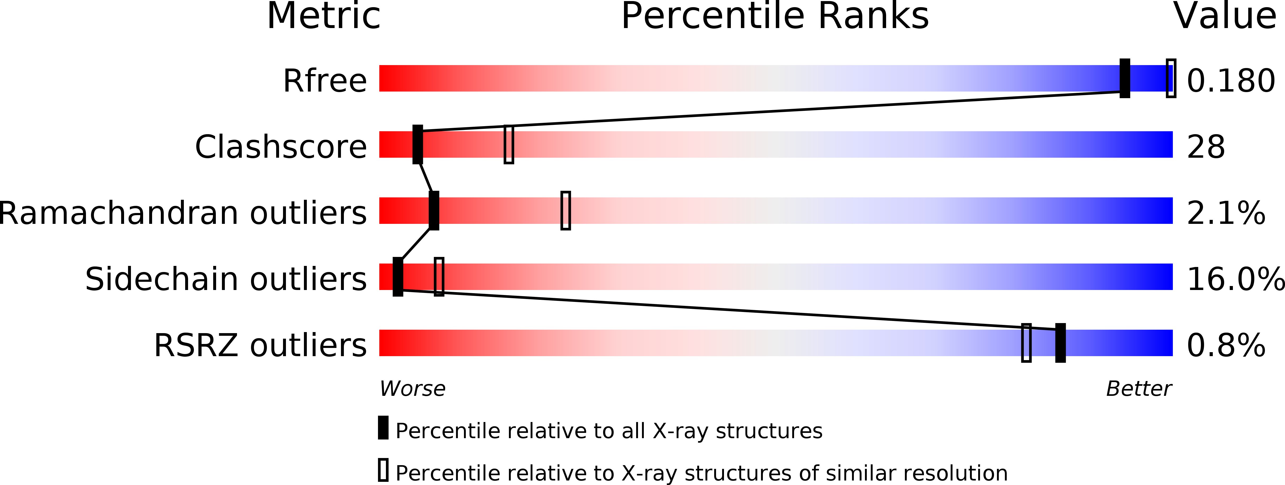
2XJ9 image
Deposition Date 2010-07-02
Release Date 2011-07-13
Last Version Date 2023-12-20
Entry Detail
PDB ID:
2XJ9
Keywords:
Title:
Dimer Structure of the bacterial cell division regulator MipZ
Biological Source:
Source Organism:
Host Organism:
Method Details:
Experimental Method:
Resolution:
2.80 Å
R-Value Free:
0.23
R-Value Work:
0.18
R-Value Observed:
0.18
Space Group:
P 41
Macromolecular Entities
Structures with similar UniProt ID
Protein Blast
Polymer Type:polypeptide(L)
Molecule:MIPZ
Mutagens:YES
Chain IDs:A, B
Chain Length:286
Number of Molecules:2
Biological Source:CAULOBACTER CRESCENTUS
Primary Citation
Localized Dimerization and Nucleoid Binding Drive Gradient Formation by the Bacterial Cell Division Inhibitor Mipz.
Mol.Cell 46 245 ? (2012)
PMID: 22483621 DOI: 10.1016/J.MOLCEL.2012.03.004

Abstact

Protein gradients play a central role in the spatial organization of cells, but the mechanisms of their formation are incompletely understood. This study analyzes the determinants responsible for establishing bipolar gradients of the ATPase MipZ, a key regulator of division site placement in Caulobacter crescentus. We have solved the crystal structure of MipZ in different nucleotide states, dissected its ATPase cycle, and investigated its interaction with FtsZ, ParB, and the nucleoid. Our results suggest that the polar ParB complexes locally stimulate the formation of ATP-bound MipZ dimers, which are then retained near the cell poles through association with chromosomal DNA. Due to their intrinsic ATPase activity, dimers eventually dissociate into freely diffusible monomers that undergo spontaneous nucleotide exchange and are recaptured by ParB. These findings clarify the molecular function of a conserved gradient-forming system and reveal mechanistic principles that might be commonly used to sustain protein gradients within cells.

Legend

Protein

Chemical

Disease

Primary Citation of related structures