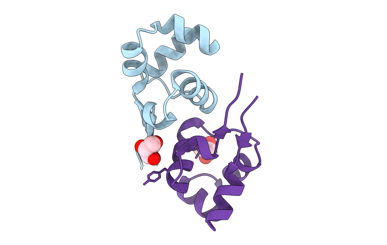
2XJ3 image
Entry Detail
PDB ID:
2XJ3
Title:
High resolution structure of the T55C mutant of CylR2.
Biological Source:
Source Organism:
Host Organism:
PDB Version:
Deposition Date:
2010-07-02
Release Date:
2011-02-09
Method Details:
Experimental Method:
Resolution:
1.23 Å
R-Value Free:
0.18
R-Value Observed:
0.15
Space Group:
P 41
Macromolecular Entities
Polymer Type:polypeptide(L)
Description:CYLR2 SYNONYM CYTOLYSIN REPRESSOR 2
Mutations:YES
Chain IDs:A, B
Chain Length:66
Number of Molecules:2
Biological Source:ENTEROCOCCUS FAECALIS
Ligand Molecules
Primary Citation
Integrated Analysis of the Conformation of a Protein-Linked Spin Label by Crystallography, Epr and NMR Spectroscopy.
J.Biomol.NMR 49 111 ? (2011)
PMID: 21271275 DOI: 10.1007/S10858-011-9471-Y

Abstact

Long-range structural information derived from paramagnetic relaxation enhancement observed in the presence of a paramagnetic nitroxide radical is highly useful for structural characterization of globular, modular and intrinsically disordered proteins, as well as protein-protein and protein-DNA complexes. Here we characterized the conformation of a spin-label attached to the homodimeric protein CylR2 using a combination of X-ray crystallography, electron paramagnetic resonance (EPR) and NMR spectroscopy. Close agreement was found between the conformation of the spin label observed in the crystal structure with interspin distances measured by EPR and signal broadening in NMR spectra, suggesting that the conformation seen in the crystal structure is also preferred in solution. In contrast, conformations of the spin label observed in crystal structures of T4 lysozyme are not in agreement with the paramagnetic relaxation enhancement observed for spin-labeled CylR2 in solution. Our data demonstrate that accurate positioning of the paramagnetic center is essential for high-resolution structure determination.

Legend

Protein

Chemical

Disease

Primary Citation of related structures