2XHZ image
Deposition Date 2010-06-24
Release Date 2011-01-26
Last Version Date 2023-12-20
Entry Detail
PDB ID:
2XHZ
Keywords:
Title:
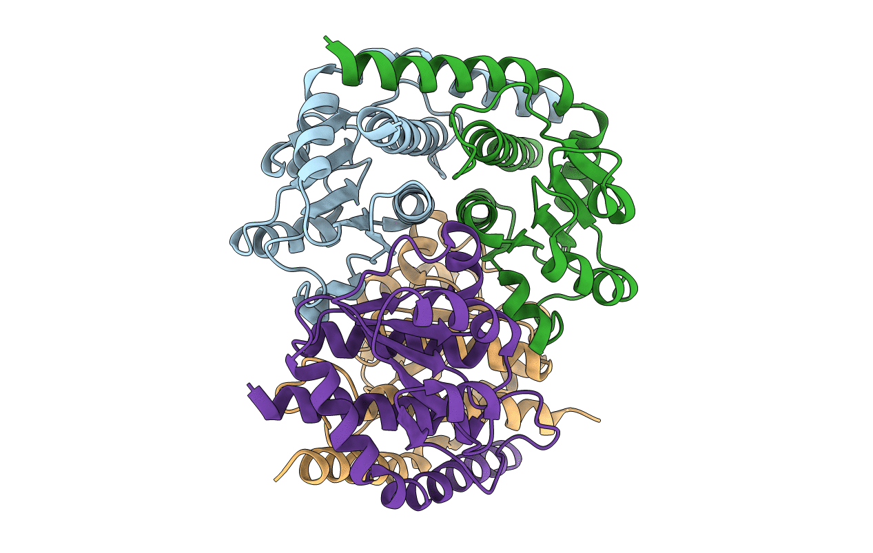
Probing the active site of the sugar isomerase domain from E. coli arabinose-5-phosphate isomerase via X-ray crystallography
Biological Source:
Source Organism:
ESCHERICHIA COLI (Taxon ID: 562)
Host Organism:
Method Details:
Experimental Method:
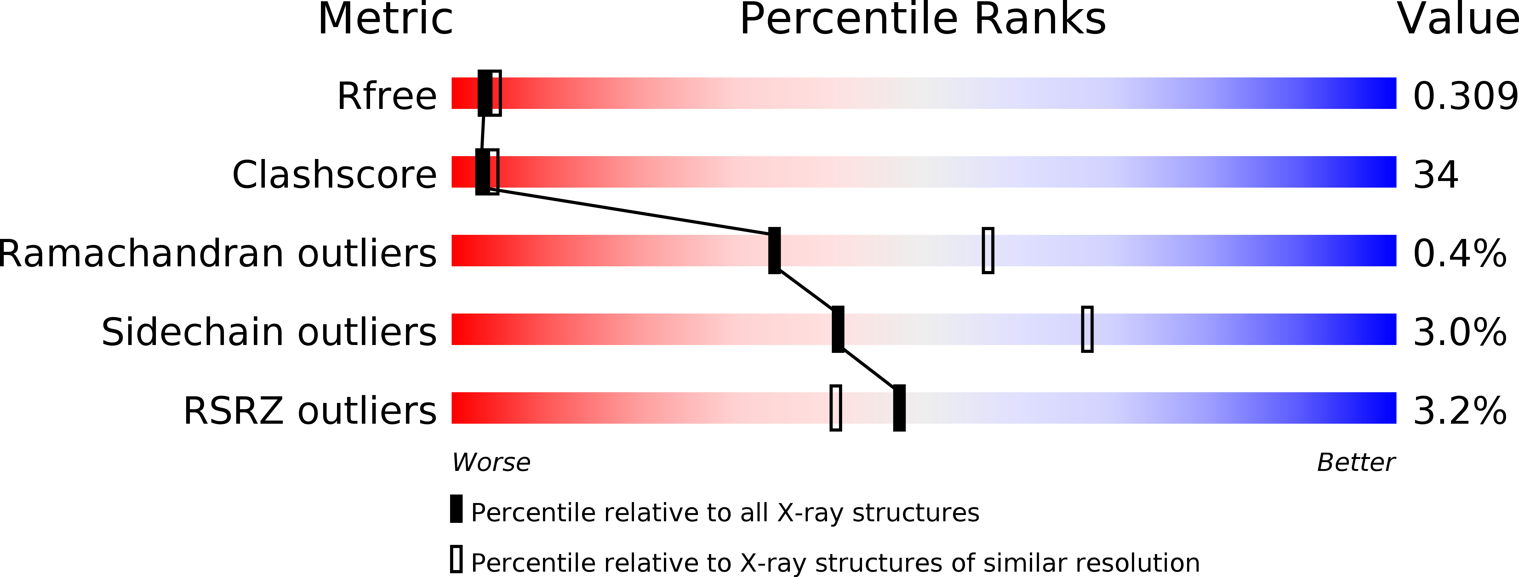
Resolution:
2.60 Å
R-Value Free:
0.30
R-Value Work:
0.26
R-Value Observed:
0.26
Space Group:
P 1 21 1
Macromolecular Entities
Structures with similar UniProt ID
Protein Blast
Polymer Type:polypeptide(L)
Molecule:ARABINOSE 5-PHOSPHATE ISOMERASE
Gene (Uniprot):kdsD
Mutagens:YES
Chain IDs:A, B, C, D
Chain Length:183
Number of Molecules:4
Biological Source:ESCHERICHIA COLI
Primary Citation
Probing the Active Site of the Sugar Isomerase Domain from E. Coli Arabinose-5-Phosphate Isomerase Via X-Ray Crystallography.
Protein Sci. 19 2430 ? (2010)
PMID: 20954237 DOI: 10.1002/PRO.525

Abstact

Lipopolysaccharide (LPS) biosynthesis represents an underexploited target pathway for novel antimicrobial development to combat the emergence of multidrug-resistant bacteria. A key player in LPS synthesis is the enzyme D-arabinose-5-phosphate isomerase (API), which catalyzes the reversible isomerization of D-ribulose-5-phosphate to D-arabinose-5-phosphate, a precursor of 3-deoxy-D-manno-octulosonate that is an essential residue of the LPS inner core. API is composed of two main domains: an N-terminal sugar isomerase domain (SIS) and a pair of cystathionine-β-synthase domains of unknown function. As the three-dimensional structure of an enzyme is a prerequisite for the rational development of novel inhibitors, we present here the crystal structure of the SIS domain of a catalytic mutant (K59A) of E. coli D-arabinose-5-phosphate isomerase at 2.6-Å resolution. Our structural analyses and comparisons made with other SIS domains highlight several potentially important active site residues. In particular, the crystal structure allowed us to identify a previously unpredicted His residue (H88) located at the mouth of the active site cavity as a possible catalytic residue. On the basis of such structural data, subsequently supported by biochemical and mutational experiments, we confirm the catalytic role of H88, which appears to be a generally conserved residue among two-domain isomerases.

Legend

Protein

Chemical

Disease

Primary Citation of related structures