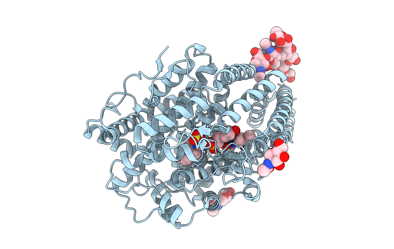
2XHM image
Deposition Date 2010-06-18
Release Date 2010-07-14
Last Version Date 2024-10-16
Entry Detail
PDB ID:
2XHM
Keywords:
Title:
Crystal structure of AnCE-K26 complex
Biological Source:
Source Organism:
Host Organism:
Method Details:
Experimental Method:
Resolution:
1.96 Å
R-Value Free:
0.21
R-Value Work:
0.19
R-Value Observed:
0.19
Space Group:
H 3
Macromolecular Entities
Polymer Type:polypeptide(L)
Molecule:ANGIOTENSIN CONVERTING ENZYME
Gene (Uniprot):Ance
Chain IDs:A
Chain Length:598
Number of Molecules:1
Biological Source:DROSOPHILA MELANOGASTER
Modified Residue
Compound ID Chain ID Parent Comp ID Details 2D Image
ASN A ASN GLYCOSYLATION SITE
Primary Citation
Crystal Structure of a Phosphonotripeptide K-26 in Complex with Angiotensin Converting Enzyme Homologue (Ance) from Drosophila Melanogaster.
Biochem.Biophys.Res.Commun. 398 532 ? (2010)
PMID: 20599761 DOI: 10.1016/J.BBRC.2010.06.113

Abstact

Angiotensin-I converting enzyme (ACE, a zinc dependent dipeptidyl carboxypeptidase) is a major target of drugs due to its role in the modulation of blood pressure and cardiovascular disorders. Here we present a crystal structure of AnCE (an ACE homologue from Drosophila melanogaster with a single enzymatic domain) in complex with a natural product-phosphonotripeptide, K-26 at 1.96A resolution. The inhibitor binds exclusively in the S(1) and S(2) binding pockets of AnCE (coordinating the zinc ion) through ionic and hydrogen bond interactions. A detailed structural comparison of AnCE.K-26 complex with individual domains of human somatic ACE provides useful information for further exploration of ACE inhibitor pharmacophores involving phosphonic acids.

Legend

Protein

Chemical

Disease

Primary Citation of related structures