Deposition Date
2010-06-08
Release Date
2010-08-25
Last Version Date
2023-12-20
Entry Detail
PDB ID:
2XH0
Keywords:
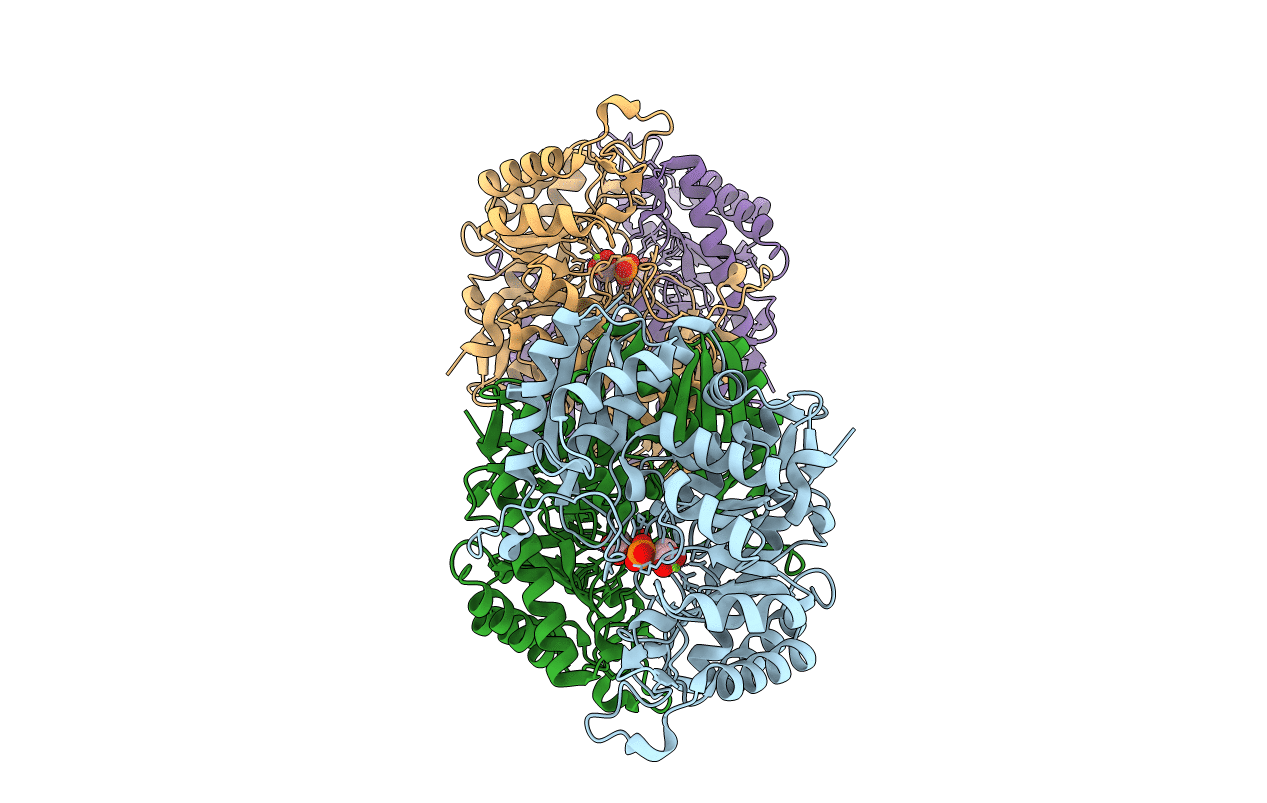
Title:
Engineering the enolase active site pocket: Crystal structure of the S39N Q167K D321R mutant of yeast enolase 1
Biological Source:
Source Organism(s):
SACCHAROMYCES CEREVISIAE (Taxon ID: 4932)
Expression System(s):
Method Details:
Experimental Method:
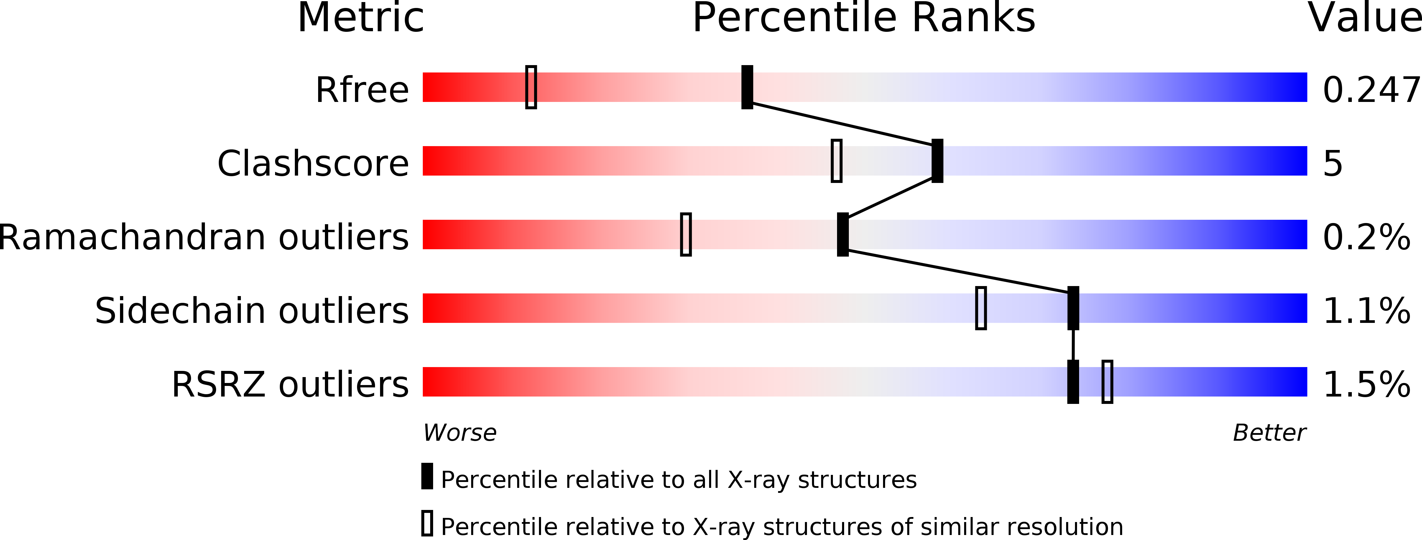
Resolution:
1.70 Å
R-Value Free:
0.24
R-Value Work:
0.19
R-Value Observed:
0.19
Space Group:
P 1