2XG7 image
Deposition Date 2010-05-31
Release Date 2010-10-13
Last Version Date 2024-10-09
Entry Detail
PDB ID:
2XG7
Title:
Crystal Structure of BST2-Tetherin Ectodomain expressed in HEK293T cells
Biological Source:
Source Organism(s):
HOMO SAPIENS (Taxon ID: 9606)
Expression System(s):
Method Details:
Experimental Method:
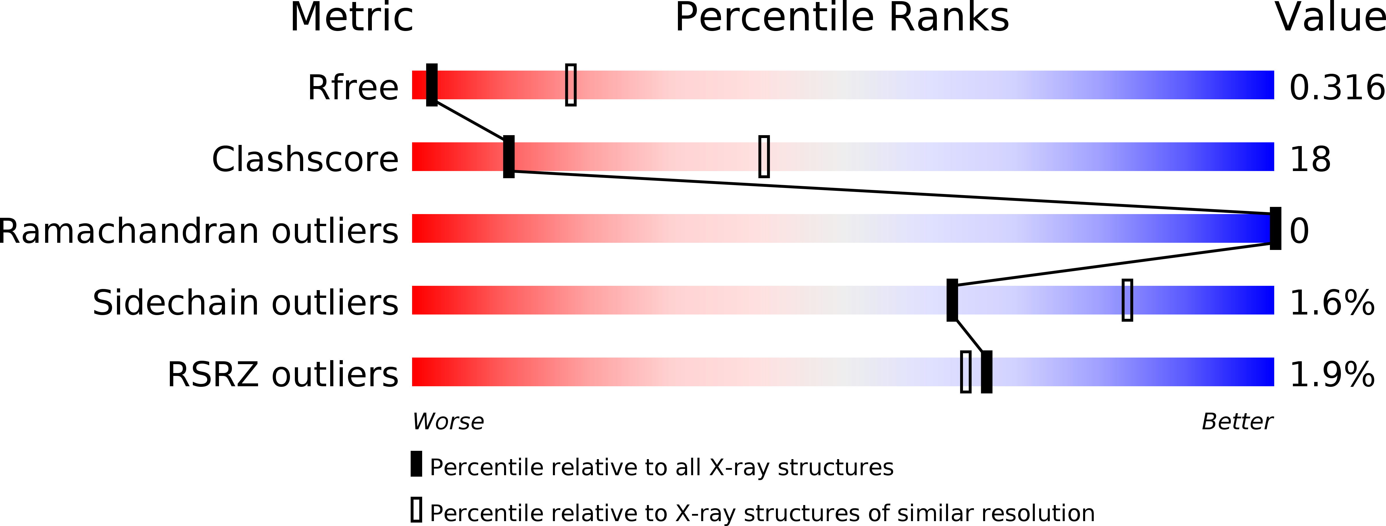
Resolution:
3.45 Å
R-Value Free:
0.29
R-Value Work:
0.26
R-Value Observed:
0.27
Space Group:
P 21 21 21
Macromolecular Entities
Structures with similar UniProt ID
Protein Blast
Polymer Type:polypeptide(L)
Molecule:BONE MARROW STROMAL ANTIGEN 2
Gene (Uniprot):BST2
Chain IDs:A, B (auth: C)
Chain Length:103
Number of Molecules:2
Biological Source:HOMO SAPIENS
Modified Residue
Compound ID Chain ID Parent Comp ID Details 2D Image
ASN A ASN GLYCOSYLATION SITE
Ligand Molecules
Primary Citation
Structural and Functional Studies on the Extracellular Domain of Bst2/Tetherin in Reduced and Oxidized Conformations.
Proc.Natl.Acad.Sci.USA 107 17951 ? (2010)
PMID: 20880831 DOI: 10.1073/PNAS.1008206107

Abstact

HIV-1 and other enveloped viruses can be restricted by a host cellular protein called BST2/tetherin that prevents release of budded viruses from the cell surface. Mature BST2 contains a small cytosolic region, a predicted transmembrane helix, and an extracellular domain with a C-terminal GPI anchor. To advance understanding of BST2 function, we have determined a 2.6 Å crystal structure of the extracellular domain of the bacterially expressed recombinant human protein, residues 47-152, under reducing conditions. The structure forms a single long helix that associates as a parallel dimeric coiled coil over its C-terminal two-thirds, while the N-terminal third forms an antiparallel four-helix bundle with another dimer, creating a global tetramer. We also report the 3.45 Å resolution structure of BST2(51-151) prepared by expression as a secreted protein in HEK293T cells. This oxidized construct forms a dimer in the crystal that is superimposable with the reduced protein over the C-terminal two-thirds of the molecule, and its N terminus suggests pronounced flexibility. Hydrodynamic data demonstrated that BST2 formed a stable tetramer under reducing conditions and a dimer when oxidized to form disulfide bonds. A mutation that selectively disrupted the tetramer (L70D) increased protein expression modestly but only reduced antiviral activity by approximately threefold. Our data raise the possibility that BST2 may function as a tetramer at some stage, such as during trafficking, and strongly support a model in which the primary functional state of BST2 is a parallel disulfide-bound coiled coil that displays flexibility toward its N terminus.

Legend

Protein

Chemical

Disease

Primary Citation of related structures
Feedback Form
Name
Email
Institute
Feedback