2XFM image
Deposition Date 2010-05-27
Release Date 2011-01-26
Last Version Date 2024-05-15
Entry Detail
PDB ID:
2XFM
Keywords:
Title:
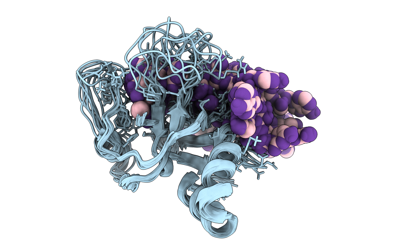
Complex structure of the MIWI Paz domain bound to methylated single stranded RNA
Biological Source:
Source Organism:
Host Organism:
Method Details:
Experimental Method:
Conformers Calculated:
100
Conformers Submitted:
10
Selection Criteria:
LOWEST ENERGY
Macromolecular Entities
Polymer Type:polypeptide(L)
Molecule:PIWI-LIKE PROTEIN 1
Gene (Uniprot):Piwil1
Chain IDs:A
Chain Length:150
Number of Molecules:1
Biological Source:MUS MUSCULUS
Polymer Type:polyribonucleotide
Molecule:5'-R(*AP*CP*CP*GP*AP*CP*UP*(OMU)P)-3'
Chain IDs:B
Chain Length:8
Number of Molecules:1
Biological Source:SYNTHETIC CONSTRUCT
Modified Residue
Compound ID Chain ID Parent Comp ID Details 2D Image
OMU B U O2'-METHYLURIDINE 5'-MONOPHOSPHATE
Ligand Molecules
Primary Citation
Recognition of 2'-O-Methylated 3'-End of Pirna by the Paz Domain of a Piwi Protein.
Structure 19 172 ? (2011)
PMID: 21237665 DOI: 10.1016/J.STR.2010.11.015

Abstact

Piwi proteins are germline-specific Argonautes that associate with small RNAs called Piwi-interacting RNAs (piRNAs), and together with these RNAs are implicated in transposon silencing. The PAZ domain of Argonaute proteins recognizes the 3'-end of the RNA, which in the case of piRNAs is invariably modified with a 2'-O-methyl group. Here, we present the solution structure of the PAZ domain from the mouse Piwi protein, MIWI, in complex with an 8-mer piRNA mimic. The methyl group is positioned in a hydrophobic cavity made of conserved amino acids from strand β7 and helix α3, where it is contacted by the side chain of methionine-382. Our structure is similar to that of Ago-PAZ, but subtle differences illustrate how the PAZ domain has evolved to accommodate distinct 3' ends from a variety of RNA substrates.

Legend

Protein

Chemical

Disease

Primary Citation of related structures