2XFH image
Deposition Date 2010-05-24
Release Date 2010-09-29
Last Version Date 2023-12-20
Entry Detail
PDB ID:
2XFH
Keywords:
Title:
Structure of cytochrome P450 EryK cocrystallized with inhibitor clotrimazole.
Biological Source:
Source Organism(s):
Expression System(s):
Method Details:
Experimental Method:
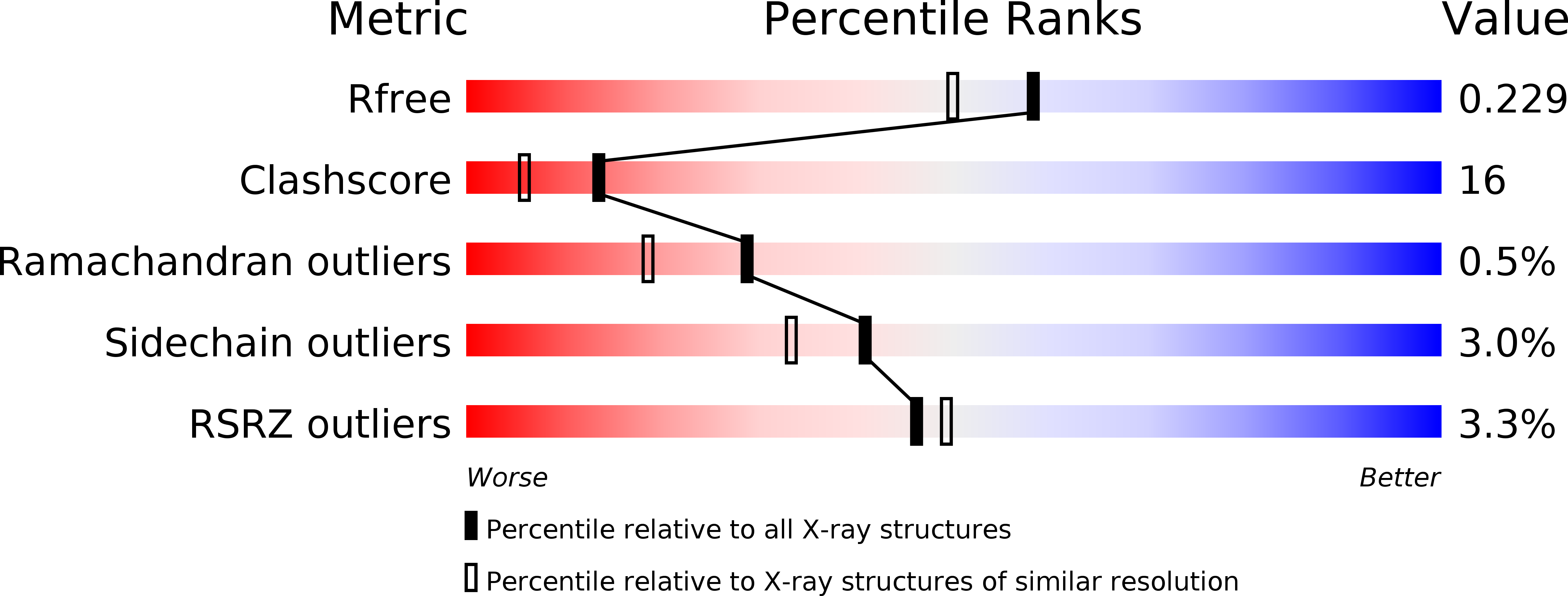
Resolution:
1.90 Å
R-Value Free:
0.23
R-Value Work:
0.18
R-Value Observed:
0.18
Space Group:
P 1
Macromolecular Entities
Structures with similar UniProt ID
Protein Blast
Polymer Type:polypeptide(L)
Molecule:ERYTHROMYCIN B/D C-12 HYDROXYLASE
Gene (Uniprot):eryK
Mutagens:YES
Chain IDs:A
Chain Length:411
Number of Molecules:1
Biological Source:SACCHAROPOLYSPORA ERYTHRAEA
Primary Citation
Azole Drugs Trap Cytochrome P450 Eryk in Alternative Conformational States.
Biochemistry 49 9199 ? (2010)
PMID: 20845962 DOI: 10.1021/BI101062V

Abstact

EryK is a bacterial cytochrome P450 that catalyzes the last hydroxylation occurring during the biosynthetic pathway of erythromycin A in Streptomyces erythraeus. We report the crystal structures of EryK in complex with two widely used azole inhibitors: ketoconazole and clotrimazole. Both of these ligands use their imidazole moiety to coordinate the heme iron of P450s. Nevertheless, because of the different chemical and structural properties of their N1-substituent group, ketoconazole and clotrimazole trap EryK, respectively, in a closed and in an open conformation that resemble the two structures previously described for the ligand-free EryK. Indeed, ligands induce a distortion of the internal helix I that affects the accessibility of the binding pocket by regulating the kink of the external helix G via a network of interactions that involves helix F. The data presented thus constitute an example of how a cytochrome P450 may be selectively trapped in different conformational states by inhibitors.

Legend

Protein

Chemical

Disease

Primary Citation of related structures
Feedback Form
Name
Email
Institute
Feedback