2XEX image
Deposition Date 2010-05-19
Release Date 2010-07-14
Last Version Date 2023-12-20
Entry Detail
PDB ID:
2XEX
Keywords:
Title:
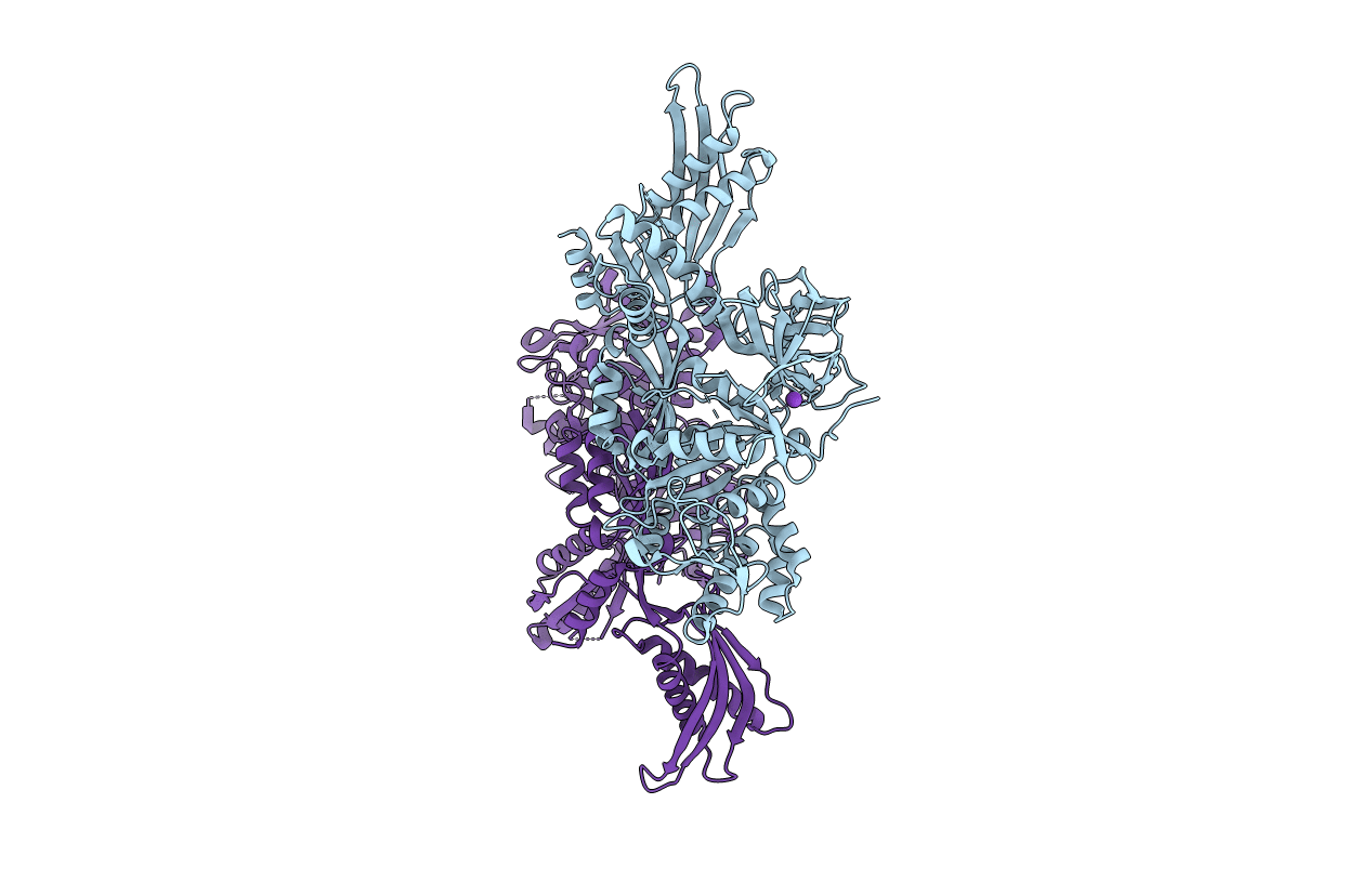
crystal structure of Staphylococcus aureus elongation factor G
Biological Source:
Source Organism:
Host Organism:
Method Details:
Experimental Method:
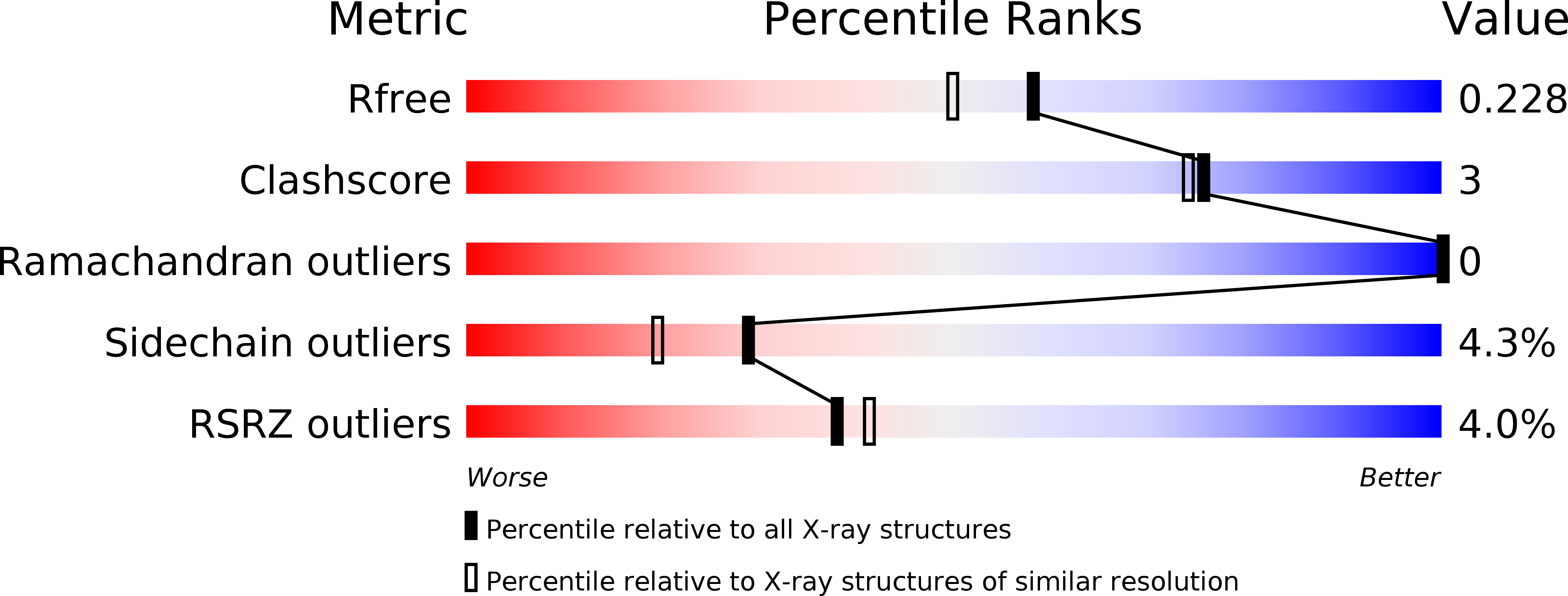
Resolution:
1.90 Å
R-Value Free:
0.22
R-Value Work:
0.18
R-Value Observed:
0.18
Space Group:
P 1 21 1
Macromolecular Entities
Structures with similar UniProt ID
Protein Blast
Polymer Type:polypeptide(L)
Molecule:ELONGATION FACTOR G
Gene (Uniprot):fusA
Chain IDs:A, B
Chain Length:693
Number of Molecules:2
Biological Source:STAPHYLOCOCCUS AUREUS
Primary Citation
Staphylococcus Aureus Elongation Factor G - Structure and Analysis of a Target for Fusidic Acid.
FEBS J. 277 3789 ? (2010)
PMID: 20718859 DOI: 10.1111/J.1742-4658.2010.07780.X

Abstact

Fusidic acid (FA) is a bacteriostatic antibiotic that locks elongation factor G (EF-G) on the ribosome in a post-translocational state. It is used clinically against Gram-positive bacteria such as pathogenic strains of Staphylococcus aureus, but no structural information has been available for EF-G from these species. We have solved the apo crystal structure of EF-G from S. aureus to 1.9 Å resolution. This structure shows a dramatically different overall conformation from previous structures of EF-G, although the individual domains are highly similar. Between the different structures of free or ribosome-bound EF-G, domains III-V move relative to domains I-II, resulting in a displacement of the tip of domain IV relative to domain G. In S. aureus EF-G, this displacement is about 25 Å relative to structures of Thermus thermophilus EF-G in a direction perpendicular to that in previous observations. Part of the switch I region (residues 46-56) is ordered in a helix, and has a distinct conformation as compared with structures of EF-Tu in the GDP and GTP states. Also, the switch II region shows a new conformation, which, as in other structures of free EF-G, is incompatible with FA binding. We have analysed and discussed all known fusA-based fusidic acid resistance mutations in the light of the new structure of EF-G from S. aureus, and a recent structure of T. thermophilus EF-G in complex with the 70S ribosome with fusidic acid [Gao YG et al. (2009) Science326, 694-699]. The mutations can be classified as affecting FA binding, EF-G-ribosome interactions, EF-G conformation, and EF-G stability.

Legend

Protein

Chemical

Disease

Primary Citation of related structures