2XDF image
Deposition Date 2010-04-30
Release Date 2010-09-22
Last Version Date 2024-05-15
Entry Detail
PDB ID:
2XDF
Keywords:
Title:
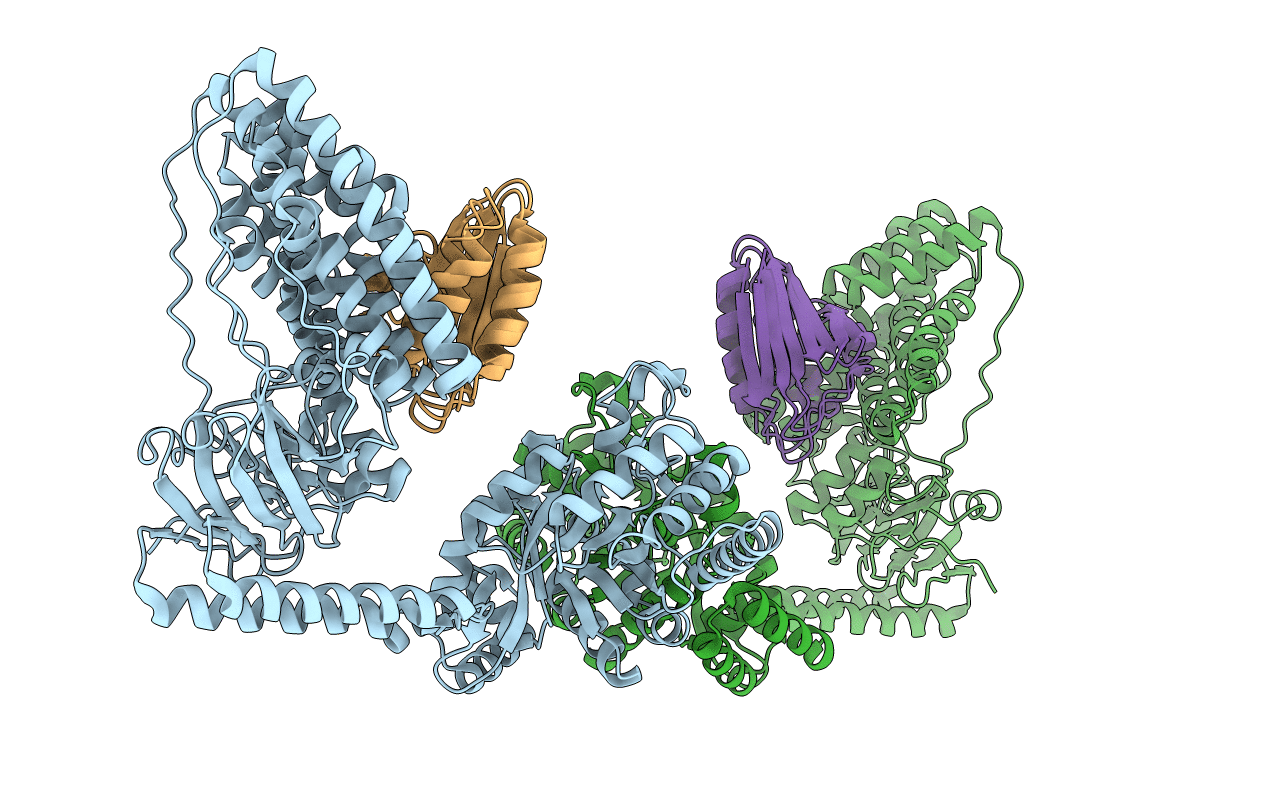
Solution Structure of the Enzyme I Dimer Complexed with HPr Using Residual Dipolar Couplings and Small Angle X-Ray Scattering
Biological Source:
Source Organism:
Host Organism:
Method Details:
Experimental Method:
Conformers Calculated:
120
Conformers Submitted:
2
Selection Criteria:
BEST EXPERIMENT FIT, AND THEN LOWEST ENERGY
Macromolecular Entities
Polymer Type:polypeptide(L)
Molecule:PHOSPHOENOLPYRUVATE-PROTEIN PHOSPHOTRANSFERASE
Gene (Uniprot):ptsI
Chain IDs:A, B
Chain Length:573
Number of Molecules:2
Biological Source:ESCHERICHIA COLI
Polymer Type:polypeptide(L)
Molecule:PHOSPHOCARRIER PROTEIN HPR
Chain IDs:C, D
Chain Length:85
Number of Molecules:2
Biological Source:ESCHERICHIA COLI
Ligand Molecules
Primary Citation
Solution Structure of the 128 kDa Enzyme I Dimer from Escherichia Coli and its 146 kDa Complex with Hpr Using Residual Dipolar Couplings and Small- and Wide-Angle X-Ray Scattering.
J.Am.Chem.Soc. 132 13026 ? (2010)
PMID: 20731394 DOI: 10.1021/JA105485B

Abstact

The solution structures of free Enzyme I (EI, ∼128 kDa, 575 × 2 residues), the first enzyme in the bacterial phosphotransferase system, and its complex with HPr (∼146 kDa) have been solved using novel methodology that makes use of prior structural knowledge (namely, the structures of the dimeric EIC domain and the isolated EIN domain both free and complexed to HPr), combined with residual dipolar coupling (RDC), small- (SAXS) and wide- (WAXS) angle X-ray scattering and small-angle neutron scattering (SANS) data. The calculational strategy employs conjoined rigid body/torsion/Cartesian simulated annealing, and incorporates improvements in calculating and refining against SAXS/WAXS data that take into account complex molecular shapes in the description of the solvent layer resulting in a better representation of the SAXS/WAXS data. The RDC data orient the symmetrically related EIN domains relative to the C(2) symmetry axis of the EIC dimer, while translational, shape, and size information is provided by SAXS/WAXS. The resulting structures are independently validated by SANS. Comparison of the structures of the free EI and the EI-HPr complex with that of the crystal structure of a trapped phosphorylated EI intermediate reveals large (∼70-90°) hinge body rotations of the two subdomains comprising the EIN domain, as well as of the EIN domain relative to the dimeric EIC domain. These large-scale interdomain motions shed light on the structural transitions that accompany the catalytic cycle of EI.

Legend

Protein

Chemical

Disease

Primary Citation of related structures