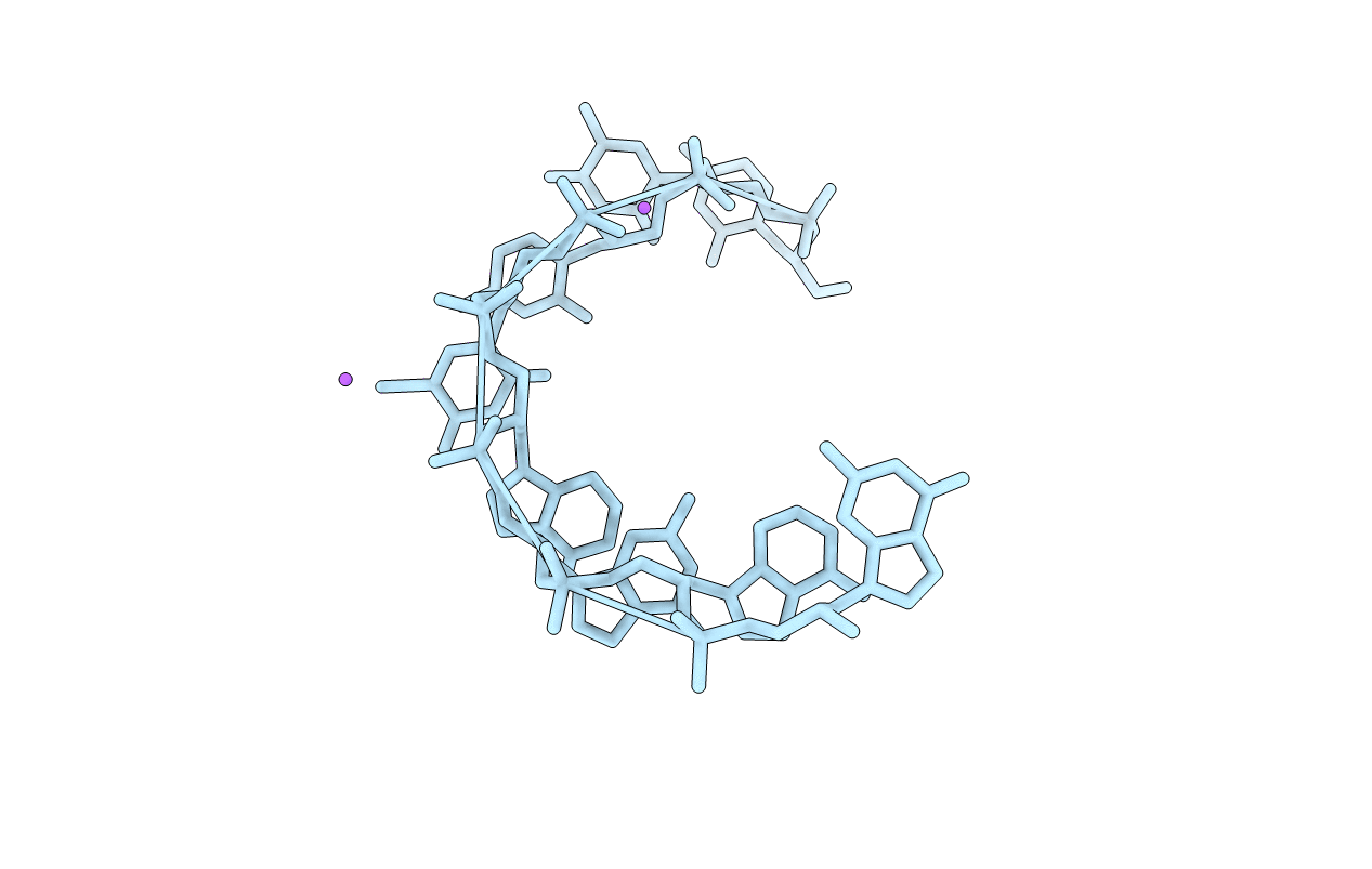
2XC6 image
Deposition Date 2010-04-16
Release Date 2011-05-04
Last Version Date 2024-06-19
Entry Detail
PDB ID:
2XC6
Keywords:
Title:
Crystal structure of the GNA 3'-CTC(Br)UAGAG-2'
Biological Source:
Source Organism:
(Taxon ID: 32630)
Method Details:
Experimental Method:
Resolution:
1.83 Å
R-Value Free:
0.27
R-Value Work:
0.23
R-Value Observed:
0.23
Space Group:
I 41 2 2
Macromolecular Entities
Polymer Type:polyribonucleotide
Molecule:GNA
Chain IDs:A
Chain Length:8
Number of Molecules:1
Biological Source:
Modified Residue
Compound ID Chain ID Parent Comp ID Details 2D Image
ZAD A A (S)-1'-(2',3'-DIHYDROXYPROPYL)-ADENINE
ZBU A U ?
ZCY A C (S)-1'-(2',3'-DIHYDROXYPROPYL)-CYTOSINE
ZGU A G (S)-1'-(2',3'-DIHYDROXYPROPYL)-GUANINE
ZTH A T (S)-1'-(2',3'-DIHYDROXYPROPYL)-THYMINE
Ligand Molecules
Primary Citation
On the Structure and Dynamics of Duplex Gna.
J.Org.Chem. 76 7964 ? (2011)
PMID: 21838272 DOI: 10.1021/JO201469B

Abstact

Glycol nucleic acid (GNA), with a nucleotide backbone comprising of just three carbons and the stereocenter derived from propylene glycol (1,2-propanediol), is a structural analog of nucleic acids with intriguing biophysical properties, such as formation of highly stable antiparallel duplexes with high Watson-Crick base pairing fidelity. Previous crystallographic studies of double stranded GNA (dsGNA) indicated two forms of backbone conformations, an elongated M-type (containing metallo-base pairs) and the condensed N-type (containing brominated base pairs). A herein presented new crystal structure of a GNA duplex at 1.8 Å resolution from self-complementary 3'-CTC(Br)UAGAG-2' GNA oligonucleotides reveals an N-type conformation with alternating gauche-anti torsions along its (O3'-C3'-C2'-O2') backbone. To elucidate the conformational state of dsGNA in solution, molecular dynamic simulations over a period of 20 ns were performed with the now available repertoire of structural information. Interestingly, dsGNA adopts conformational states in solution intermediate between experimentally observed backbone conformations: simulated dsGNA shows the all-gauche conformation characteristic of M-type GNA with the higher helical twist common to N-type GNA structures. The so far counterintuitive, smaller loss of entropy upon duplex formation as compared to DNA can be traced back to the conformational flexibility inherent to dsGNA but missing in dsDNA. Besides extensive interstrand base stacking and conformational preorganization of single strands, this flexibility contributes to the extraordinary thermal stability of GNA.

Legend

Protein

Chemical

Disease

Primary Citation of related structures