2XA8 image
Deposition Date 2010-03-30
Release Date 2011-05-11
Last Version Date 2024-10-16
Entry Detail
PDB ID:
2XA8
Keywords:
Title:
Crystal structure of the Fab domain of omalizumab at 2.41A
Biological Source:
Source Organism:
HOMO SAPIENS (Taxon ID: 9606)
Method Details:
Experimental Method:
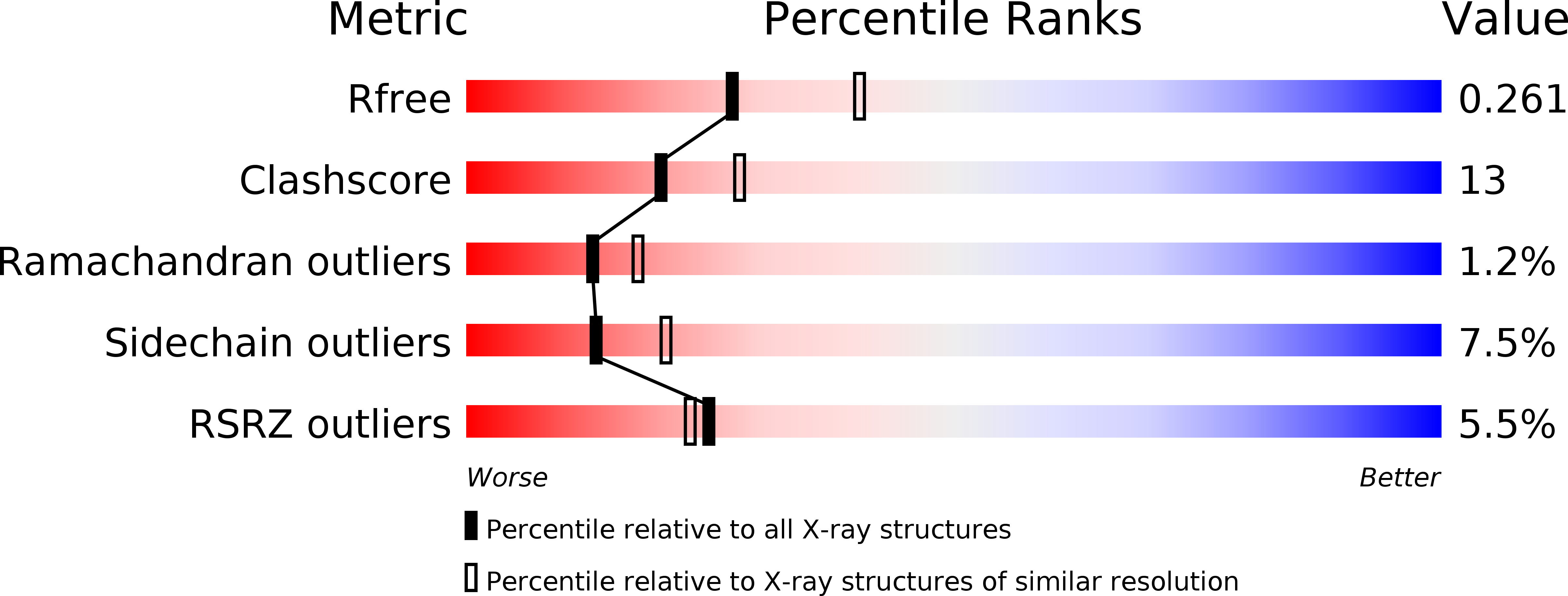
Resolution:
2.42 Å
R-Value Free:
0.26
R-Value Work:
0.22
R-Value Observed:
0.22
Space Group:
P 21 21 21
Macromolecular Entities
Polymer Type:polypeptide(L)
Molecule:OMALIZUMAB HEAVY CHAIN
Chain IDs:A (auth: H)
Chain Length:219
Number of Molecules:1
Biological Source:HOMO SAPIENS
Polymer Type:polypeptide(L)
Molecule:OMALIZUMAB LIGHT CHAIN
Chain IDs:B (auth: L)
Chain Length:218
Number of Molecules:1
Biological Source:HOMO SAPIENS
Primary Citation
Structural and Physical Basis for Anti-IgE Therapy.
Sci Rep 5 11581 11581 (2015)
PMID: 26113483 DOI: 10.1038/srep11581

Abstact

Omalizumab, an anti-IgE antibody, used to treat severe allergic asthma and chronic idiopathic urticaria, binds to IgE in blood or membrane-bound on B lymphocytes but not to IgE bound to its high (FcεRI) or low (CD23) affinity receptor. Mutagenesis studies indicate overlapping FcεRI and omalizumab-binding sites in the Cε3 domain, but crystallographic studies show FcεRI and CD23-binding sites that are far apart, so how can omalizumab block IgE from binding both receptors? We report a 2.42-Å omalizumab-Fab structure, a docked IgE-Fc/omalizumab-Fab structure consistent with available experimental data, and the free energy contributions of IgE residues to binding omalizumab, CD23, and FcεRI. These results provide a structural and physical basis as to why omalizumab cannot bind receptor-bound IgE and why omalizumab-bound IgE cannot bind to CD23/FcεRI. They reveal the key IgE residues and their roles in binding omalizumab, CD23, and FcεRI.

Legend

Protein

Chemical

Disease

Primary Citation of related structures