2X9O image
Deposition Date 2010-03-23
Release Date 2011-03-30
Last Version Date 2024-11-13
Entry Detail
PDB ID:
2X9O
Keywords:
Title:
STRUCTURE OF 15, 16- DIHYDROBILIVERDIN:FERREDOXIN OXIDOREDUCTASE (PebA)
Biological Source:
Source Organism(s):
Expression System(s):
Method Details:
Experimental Method:
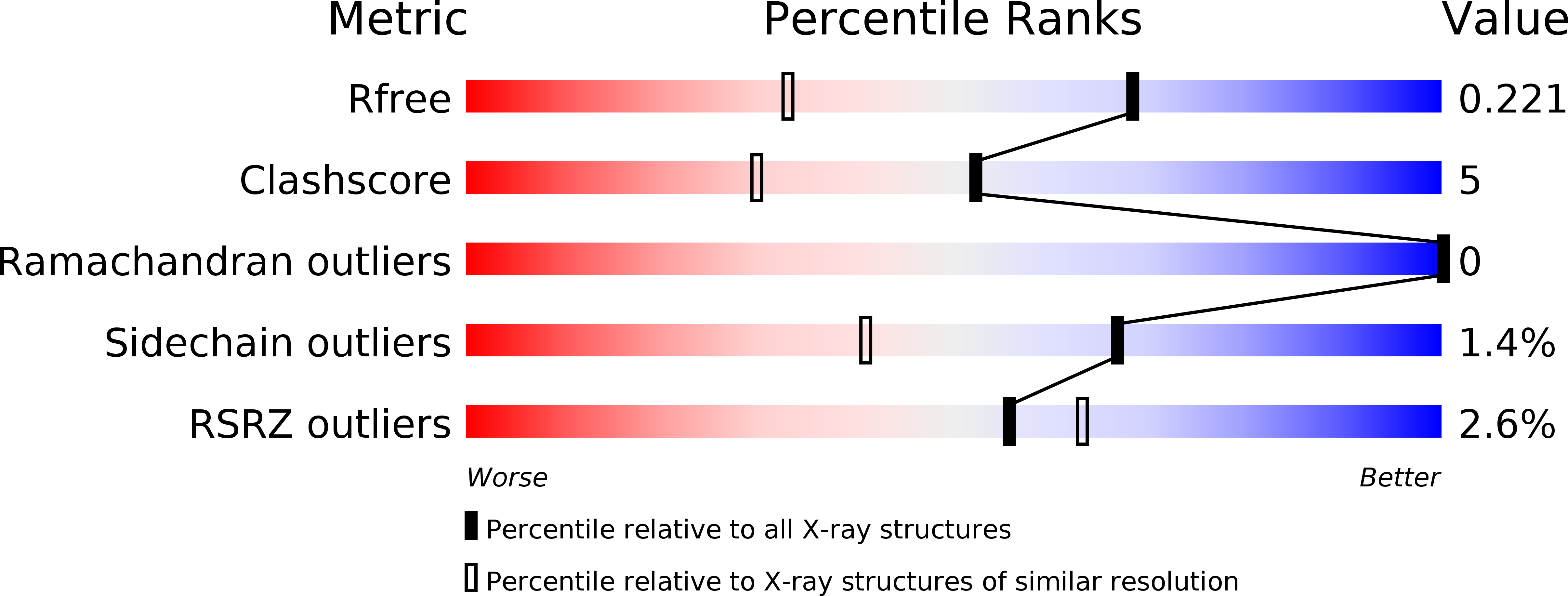
Resolution:
1.55 Å
R-Value Free:
0.22
R-Value Work:
0.17
R-Value Observed:
0.18
Space Group:
P 1 21 1
Macromolecular Entities
Structures with similar UniProt ID
Protein Blast
Polymer Type:polypeptide(L)
Molecule:15,16-DIHYDROBILIVERDIN-FERREDOXIN OXIDOREDUCTASE
Gene (Uniprot):pebA
Chain IDs:A
Chain Length:244
Number of Molecules:1
Biological Source:SYNECHOCOCCUS SP.
Modified Residue
Compound ID Chain ID Parent Comp ID Details 2D Image
MSE A MET SELENOMETHIONINE
Ligand Molecules
Primary Citation
Structural and Mechanistic Insight Into the Ferredoxin-Mediated Two-Electron Reduction of Bilins.
Biochem.J. 439 257 ? (2011)
PMID: 21729003 DOI: 10.1042/BJ20110814

Abstact

PEB (phycoerythrobilin) is one of the major open-chain tetrapyrrole molecules found in cyanobacterial light-harvesting phycobiliproteins. In these organisms, two enzymes of the ferredoxin-dependent bilin reductase family work in tandem to reduce BV (biliverdin IXα) to PEB. In contrast, a single cyanophage-encoded enzyme of the same family has been identified to catalyse the identical reaction. Using UV-visible and EPR spectroscopy we investigated the two individual cyanobacterial enzymes PebA [15,16-DHBV (dihydrobiliverdin):ferredoxin oxidoreductase] and PebB (PEB:ferredoxin oxidoreductase) showing that the two subsequent reactions catalysed by the phage enzyme PebS (PEB synthase) are clearly dissected in the cyanobacterial versions. Although a highly conserved aspartate residue is critical for both reductions, a second conserved aspartate residue is only involved in the A-ring reduction of the tetrapyrrole in PebB and PebS. The crystal structure of PebA from Synechococcus sp. WH8020 in complex with its substrate BV at a 1.55 Å (1 Å=0.1 nm) resolution revealed further insight into the understanding of enzyme evolution and function. Based on the structure it becomes obvious that in addition to the importance of certain catalytic residues, the shape of the active site and consequently the binding of the substrate highly determines the catalytic properties.

Legend

Protein

Chemical

Disease

Primary Citation of related structures
Feedback Form
Name
Email
Institute
Feedback