Deposition Date
2010-03-01
Release Date
2010-03-23
Last Version Date
2025-04-09
Entry Detail
PDB ID:
2X7K
Keywords:
Title:
The crystal structure of PPIL1 in complex with cyclosporine A suggests a binding mode for SKIP
Biological Source:
Source Organism(s):
HOMO SAPIENS (Taxon ID: 9606)
TOLYPOCLADIUM INFLATUM (Taxon ID: 29910)
TOLYPOCLADIUM INFLATUM (Taxon ID: 29910)
Expression System(s):
Method Details:
Experimental Method:
Resolution:
1.15 Å
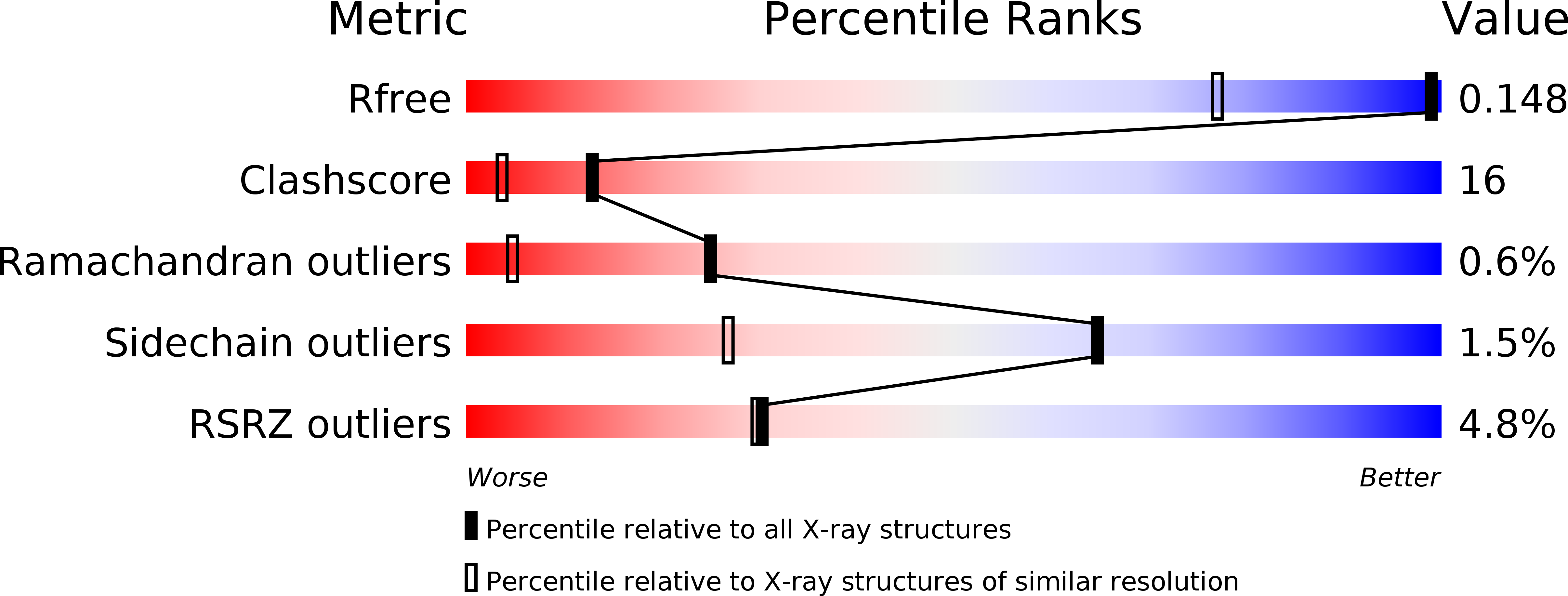
R-Value Free:
0.14
R-Value Work:
0.12
R-Value Observed:
0.12
Space Group:
P 21 21 2