2X72 image
Deposition Date 2010-02-22
Release Date 2011-03-16
Last Version Date 2024-11-13
Entry Detail
PDB ID:
2X72
Title:
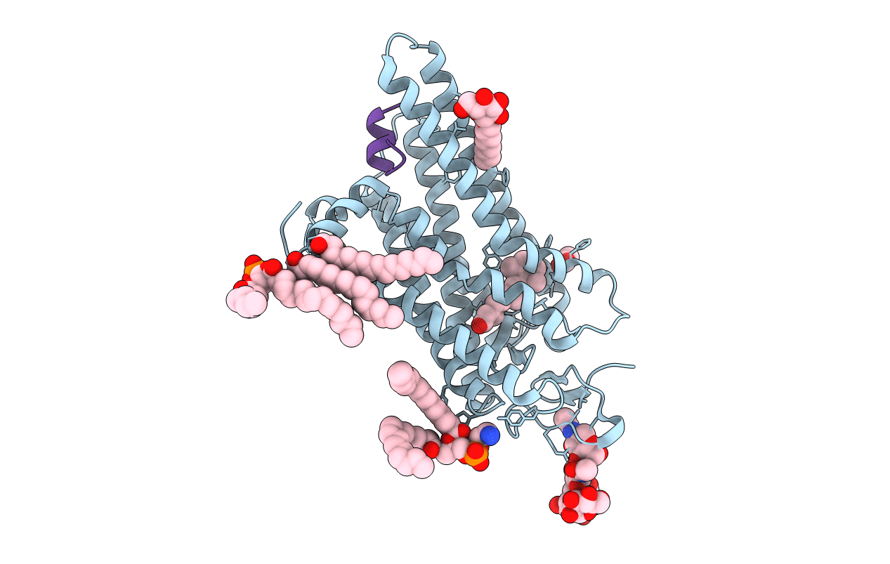
CRYSTAL STRUCTURE OF THE CONSTITUTIVELY ACTIVE E113Q,D2C,D282C RHODOPSIN MUTANT WITH BOUND GALPHACT PEPTIDE.
Biological Source:
Source Organism:
BOS TAURUS (Taxon ID: 9913)
Host Organism:
Method Details:
Experimental Method:
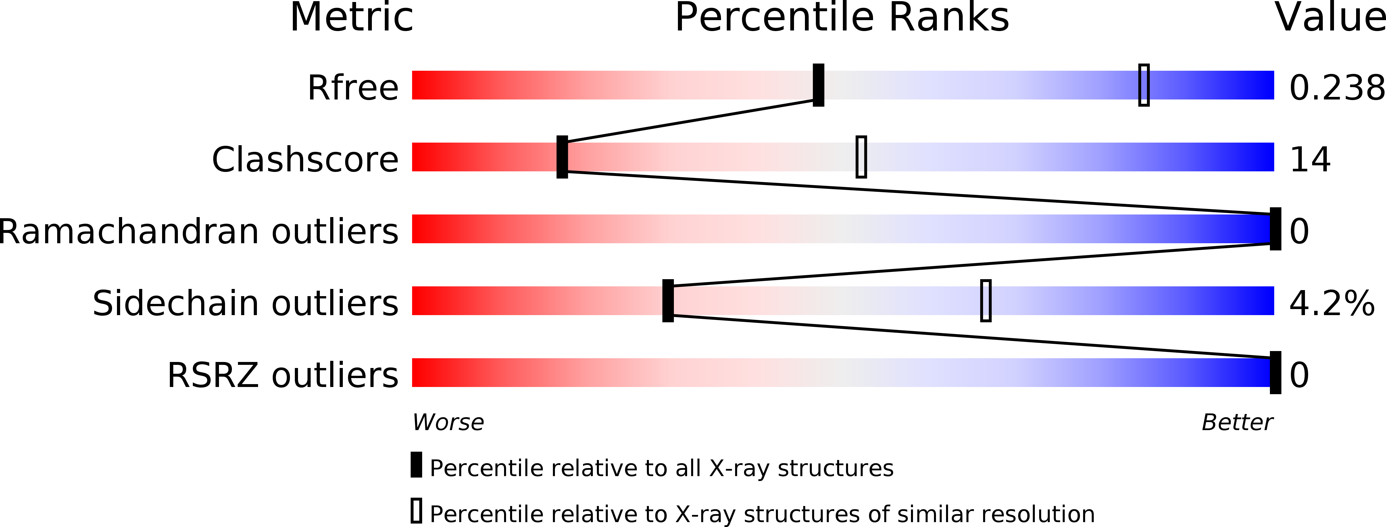
Resolution:
3.00 Å
R-Value Free:
0.24
R-Value Work:
0.21
R-Value Observed:
0.21
Space Group:
H 3 2
Macromolecular Entities
Structures with similar UniProt ID
Protein Blast
Polymer Type:polypeptide(L)
Molecule:RHODOPSIN
Gene (Uniprot):RHO
Mutagens:YES
Chain IDs:A
Chain Length:349
Number of Molecules:1
Biological Source:BOS TAURUS
Structures with similar UniProt ID
Protein Blast
Polymer Type:polypeptide(L)
Molecule:GUANINE NUCLEOTIDE-BINDING PROTEIN G(T) SUBUNIT ALPHA-1
Gene (Uniprot):GNAT1
Mutagens:YES
Chain IDs:B
Chain Length:11
Number of Molecules:1
Biological Source:BOS TAURUS
Modified Residue
Compound ID Chain ID Parent Comp ID Details 2D Image
ASN A ASN GLYCOSYLATION SITE
Primary Citation
The Structural Basis of Agonist Induced Activation in Constitutively Active Rhodopsin
Nature 471 656 ? (2011)
PMID: 21389983 DOI: 10.1038/NATURE09795

Abstact

G-protein-coupled receptors (GPCRs) comprise the largest family of membrane proteins in the human genome and mediate cellular responses to an extensive array of hormones, neurotransmitters and sensory stimuli. Although some crystal structures have been determined for GPCRs, most are for modified forms, showing little basal activity, and are bound to inverse agonists or antagonists. Consequently, these structures correspond to receptors in their inactive states. The visual pigment rhodopsin is the only GPCR for which structures exist that are thought to be in the active state. However, these structures are for the apoprotein, or opsin, form that does not contain the agonist all-trans retinal. Here we present a crystal structure at a resolution of 3 Å for the constitutively active rhodopsin mutant Glu 113 Gln in complex with a peptide derived from the carboxy terminus of the α-subunit of the G protein transducin. The protein is in an active conformation that retains retinal in the binding pocket after photoactivation. Comparison with the structure of ground-state rhodopsin suggests how translocation of the retinal β-ionone ring leads to a rotation of transmembrane helix 6, which is the critical conformational change on activation. A key feature of this conformational change is a reorganization of water-mediated hydrogen-bond networks between the retinal-binding pocket and three of the most conserved GPCR sequence motifs. We thus show how an agonist ligand can activate its GPCR.

Legend

Protein

Chemical

Disease