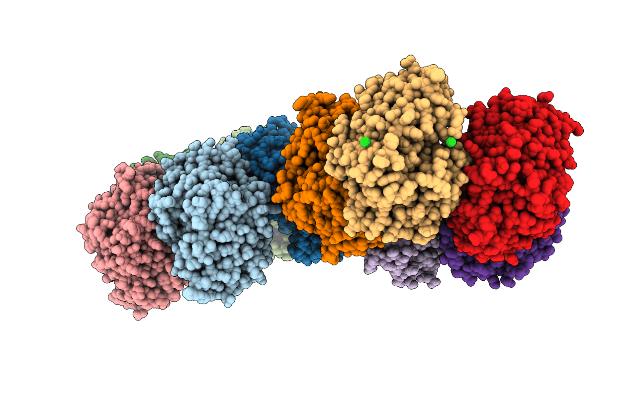
2X6T image
Deposition Date 2010-02-21
Release Date 2010-03-02
Last Version Date 2023-12-20
Entry Detail
PDB ID:
2X6T
Keywords:
Title:
AGME bound to ADP-B-mannose
Biological Source:
Source Organism:
ESCHERICHIA COLI (Taxon ID: 562)
Host Organism:
Method Details:
Experimental Method:
Resolution:
2.36 Å
R-Value Free:
0.20
R-Value Work:
0.18
R-Value Observed:
0.19
Space Group:
C 1 2 1
Macromolecular Entities
Polymer Type:polypeptide(L)
Molecule:ADP-L-GLYCERO-D-MANNO-HEPTOSE-6-EPIMERASE
Mutations:YES
Chain IDs:A, B, C, D, E, F, G, H, I, J
Chain Length:357
Number of Molecules:10
Biological Source:ESCHERICHIA COLI
Primary Citation
The Crystal Structure of the Y140F Mutant of Adp-L-Glycero-D-Manno-Heptose 6-Epimerase Bound to Adp-Beta-D-Mannose Suggests a One Base Mechanism.
Protein Sci. 19 1337 ? (2010)
PMID: 20506248 DOI: 10.1002/PRO.410

Abstact

Bacteria synthesize a wide array of unusual carbohydrate molecules, which they use in a variety of ways. The carbohydrate L-glycero-D-manno-heptose is an important component of lipopolysaccharide and is synthesized in a complex series of enzymatic steps. One step involves the epimerization at the C6'' position converting ADP-D-glycero-D-manno-heptose into ADP-L-glycero-D-manno-heptose. The enzyme responsible is a member of the short chain dehydrogenase superfamily, known as ADP-L-glycero-D-manno-heptose 6-epimerase (AGME). The structure of the enzyme was known but the arrangement of the catalytic site with respect to the substrate is unclear. We now report the structure of AGME bound to a substrate mimic, ADP-beta-D-mannose, which has the same stereochemical configuration as the substrate. The complex identifies the key residues and allows mechanistic insight into this novel enzyme.

Legend

Protein

Chemical

Disease

Primary Citation of related structures