
Deposition Date
2010-02-05
Release Date
2010-02-16
Last Version Date
2023-12-20
Entry Detail
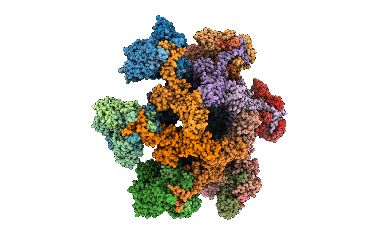
PDB ID:
2X53
Keywords:
Title:
Structure of the phage p2 baseplate in its activated conformation with Sr
Biological Source:
Source Organism(s):
LACTOCOCCUS PHAGE P2 (Taxon ID: 254252)
Expression System(s):
Method Details:
Experimental Method:
Resolution:
3.90 Å
R-Value Free:
0.24
R-Value Work:
0.22
R-Value Observed:
0.22
Space Group:
C 1 2 1
