2X3J image
Deposition Date 2010-01-25
Release Date 2011-01-19
Last Version Date 2023-12-20
Entry Detail
PDB ID:
2X3J
Keywords:
Title:
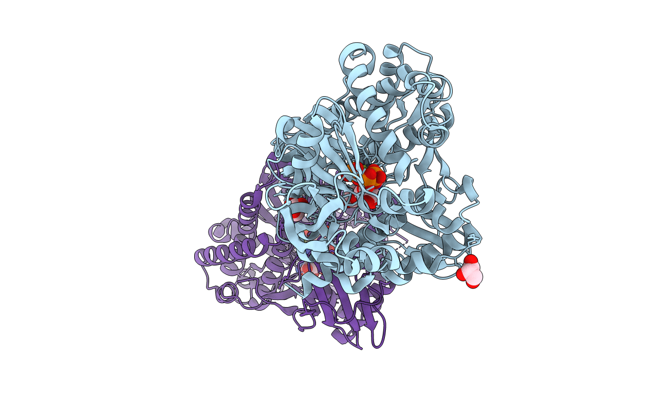
CO-COMPLEX STRUCTURE OF ACHROMOBACTIN SYNTHETASE PROTEIN D (ACSD) WITH ATP AND N-CITRYL-ETHYLENEDIAMINE FROM PECTOBACTERIUM CHRYSANTHEMI
Biological Source:
Source Organism:
Host Organism:
Method Details:
Experimental Method:
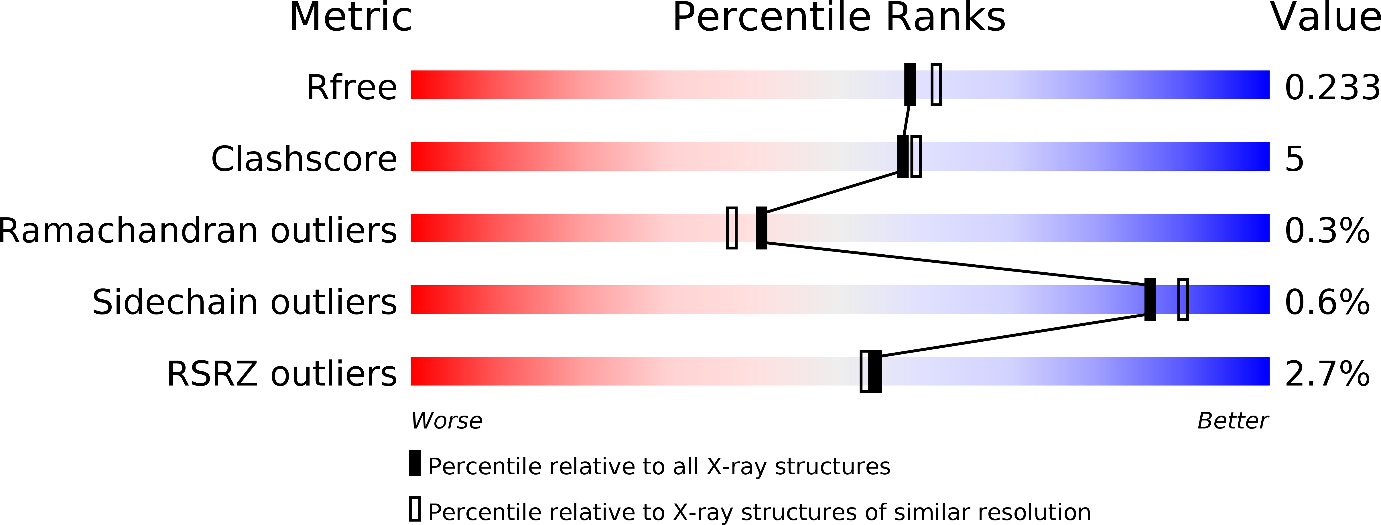
Resolution:
2.00 Å
R-Value Free:
0.23
R-Value Work:
0.18
R-Value Observed:
0.19
Space Group:
P 1
Macromolecular Entities
Structures with similar UniProt ID
Protein Blast
Polymer Type:polypeptide(L)
Molecule:ACSD
Gene (Uniprot):acsD
Chain IDs:A, B
Chain Length:620
Number of Molecules:2
Biological Source:ERWINIA CHRYSANTHEMI
Primary Citation
Structural Basis for Acyl Acceptor Specificity in the Achromobactin Biosynthetic Enzyme Acsd.
J.Mol.Biol. 412 495 ? (2011)
PMID: 21835184 DOI: 10.1016/J.JMB.2011.07.059

Abstact

Siderophores are known virulence factors, and their biosynthesis is a target for new antibacterial agents. A non-ribosomal peptide synthetase-independent siderophore biosynthetic pathway in Dickeya dadantii is responsible for production of the siderophore achromobactin. The D. dadantii achromobactin biosynthesis protein D (AcsD) enzyme has been shown to enantioselectively esterify citric acid with l-serine in the first committed step of achromobactin biosynthesis. The reaction occurs in two steps: stereospecific activation of citric acid by adenylation, followed by attack of the enzyme-bound citryl adenylate by l-serine to produce the homochiral ester. We now report a detailed characterization of the substrate profile and mechanism of the second (acyl transfer) step of AcsD enzyme. We demonstrate that the enzyme catalyzes formation of not only esters but also amides from the citryl-adenylate intermediate. We have rationalized the substrate utilization profile for the acylation reaction by determining the first X-ray crystal structure of a product complex for this enzyme class. We have identified the residues that are important for both recognition of l-serine and catalysis of ester formation. Our hypotheses were tested by biochemical analysis of various mutants, one of which shows a reversal of specificity from the wild type with respect to non-natural substrates. This change can be rationalized on the basis of our structural data. That this change in specificity is accompanied by no loss in activity suggests that AcsD and other members of the non-ribosomal peptide synthetase-independent siderophore superfamily may have biotransformation potential.

Legend

Protein

Chemical

Disease

Primary Citation of related structures
Feedback Form
Name
Email
Institute
Feedback