2X36 image
Deposition Date 2010-01-21
Release Date 2010-05-19
Last Version Date 2023-12-20
Entry Detail
PDB ID:
2X36
Keywords:
Title:
Structure of the proteolytic domain of the Human Mitochondrial Lon protease
Biological Source:
Source Organism:
HOMO SAPIENS (Taxon ID: 9606)
Host Organism:
Method Details:
Experimental Method:
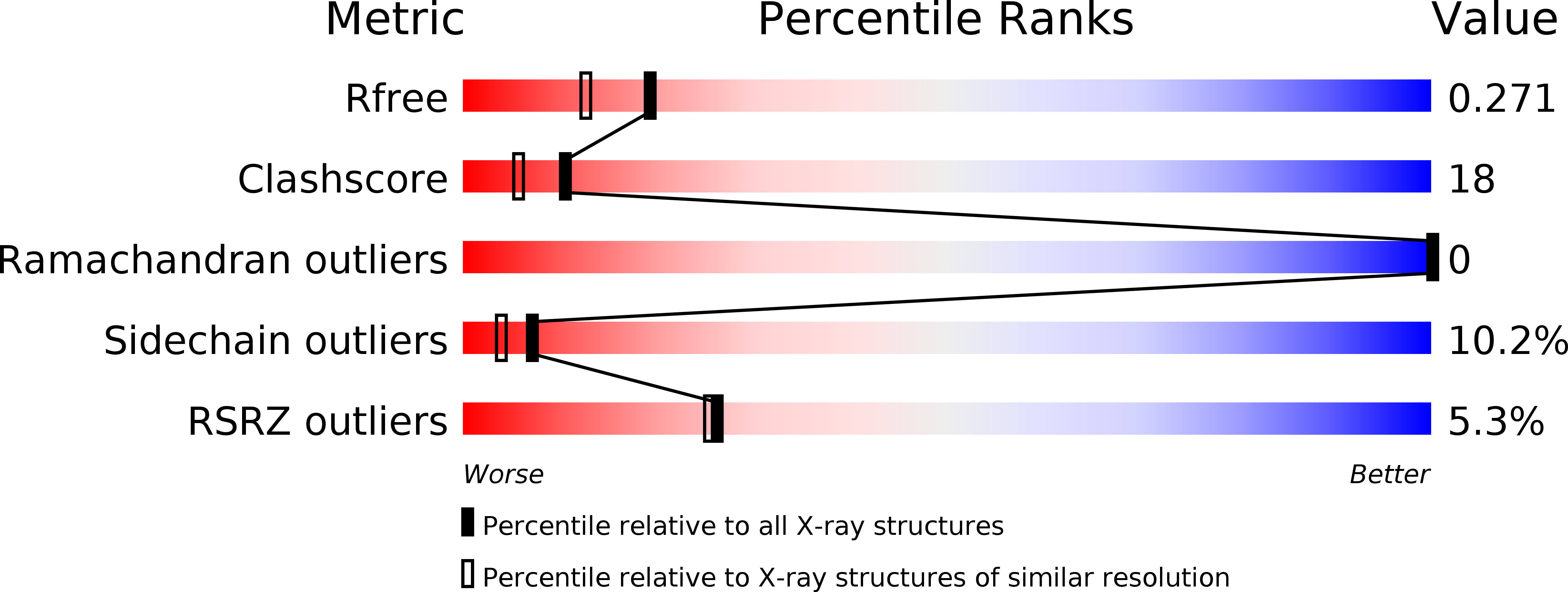
Resolution:
2.00 Å
R-Value Free:
0.23
R-Value Work:
0.19
R-Value Observed:
0.19
Space Group:
P 1 21 1
Macromolecular Entities
Structures with similar UniProt ID
Protein Blast
Polymer Type:polypeptide(L)
Molecule:LON PROTEASE HOMOLOG, MITOCHONDRIAL
Gene (Uniprot):LONP1
Chain IDs:A, B, C, D, E, F
Chain Length:207
Number of Molecules:6
Biological Source:HOMO SAPIENS
Primary Citation
Structure of the Catalytic Domain of the Human Mitochondrial Lon Protease: Proposed Relation of Oligomer Formation and Activity.
Protein Sci. 19 987 ? (2010)
PMID: 20222013 DOI: 10.1002/PRO.376

Abstact

ATP-dependent proteases are crucial for cellular homeostasis. By degrading short-lived regulatory proteins, they play an important role in the control of many cellular pathways and, through the degradation of abnormally misfolded proteins, protect the cell from a buildup of aggregates. Disruption or disregulation of mammalian mitochondrial Lon protease leads to severe changes in the cell, linked with carcinogenesis, apoptosis, and necrosis. Here we present the structure of the proteolytic domain of human mitochondrial Lon at 2 A resolution. The fold resembles those of the three previously determined Lon proteolytic domains from Escherichia coli, Methanococcus jannaschii, and Archaeoglobus fulgidus. There are six protomers in the asymmetric unit, four arranged as two dimers. The intersubunit interactions within the two dimers are similar to those between adjacent subunits of the hexameric ring of E. coli Lon, suggesting that the human Lon proteolytic domain also forms hexamers. The active site contains a 3(10) helix attached to the N-terminal end of alpha-helix 2, which leads to the insertion of Asp852 into the active site, as seen in M. jannaschii. Structural considerations make it likely that this conformation is proteolytically inactive. When comparing the intersubunit interactions of human with those of E. coli Lon taken with biochemical data leads us to propose a mechanism relating the formation of Lon oligomers with a conformational shift in the active site region coupled to a movement of a loop in the oligomer interface, converting the proteolytically inactive form seen here to the active one in the E. coli hexamer.

Legend

Protein

Chemical

Disease

Primary Citation of related structures