2X2L image
Deposition Date 2010-01-13
Release Date 2010-02-09
Last Version Date 2024-11-06
Entry Detail
PDB ID:
2X2L
Keywords:
Title:
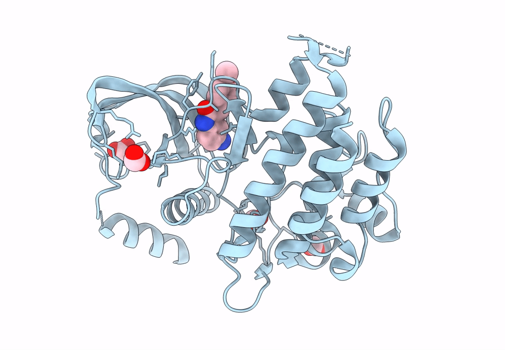
Crystal Structure of phosphorylated RET tyrosine kinase domain with inhibitor
Biological Source:
Source Organism:
HOMO SAPIENS (Taxon ID: 9606)
Host Organism:
Method Details:
Experimental Method:
Resolution:
2.00 Å
R-Value Free:
0.26
R-Value Work:
0.21
R-Value Observed:
0.21
Space Group:
C 1 2 1
Macromolecular Entities
Polymer Type:polypeptide(L)
Molecule:PROTO-ONCOGENE TYROSINE-PROTEIN KINASE RECEPTOR RET
Gene (Uniprot):RET
Chain IDs:A
Chain Length:314
Number of Molecules:1
Biological Source:HOMO SAPIENS
Modified Residue
Compound ID Chain ID Parent Comp ID Details 2D Image
PTR A TYR O-PHOSPHOTYROSINE
Primary Citation
Synthesis, structure-activity relationship and crystallographic studies of 3-substituted indolin-2-one RET inhibitors.
Bioorg. Med. Chem. 18 1482 1496 (2010)
PMID: 20117004 DOI: 10.1016/j.bmc.2010.01.011

Abstact

The synthesis, structure-activity relationships (SAR) and structural data of a series of indolin-2-one inhibitors of RET tyrosine kinase are described. These compounds were designed to explore the available space around the indolinone scaffold within RET active site. Several substitutions at different positions were tested and biochemical data were used to draw a molecular model of steric and electrostatic interactions, which can be applied to design more potent and selective RET inhibitors. The crystal structures of RET kinase domain in complex with three inhibitors were solved. All three compounds bound in the ATP pocket and formed two hydrogen bonds with the kinase hinge region. Crystallographic analysis confirmed predictions from molecular modelling and helped refine SAR results. These data provide important information for the development of indolinone inhibitors for the treatment of RET-driven cancers.

Legend

Protein

Chemical

Disease

Primary Citation of related structures