2X2I image
Deposition Date 2010-01-13
Release Date 2011-01-19
Last Version Date 2024-10-16
Entry Detail
PDB ID:
2X2I
Keywords:
Title:
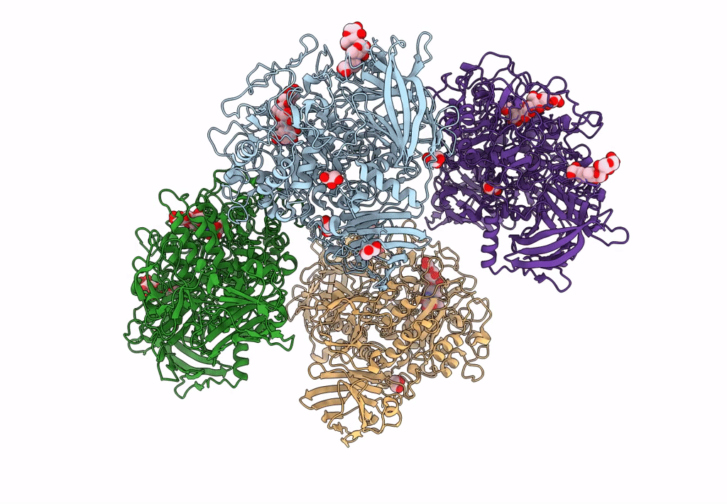
Crystal structure of the Gracilariopsis lemaneiformis alpha-1,4- glucan lyase with acarbose
Biological Source:
Source Organism:
Host Organism:
Method Details:
Experimental Method:
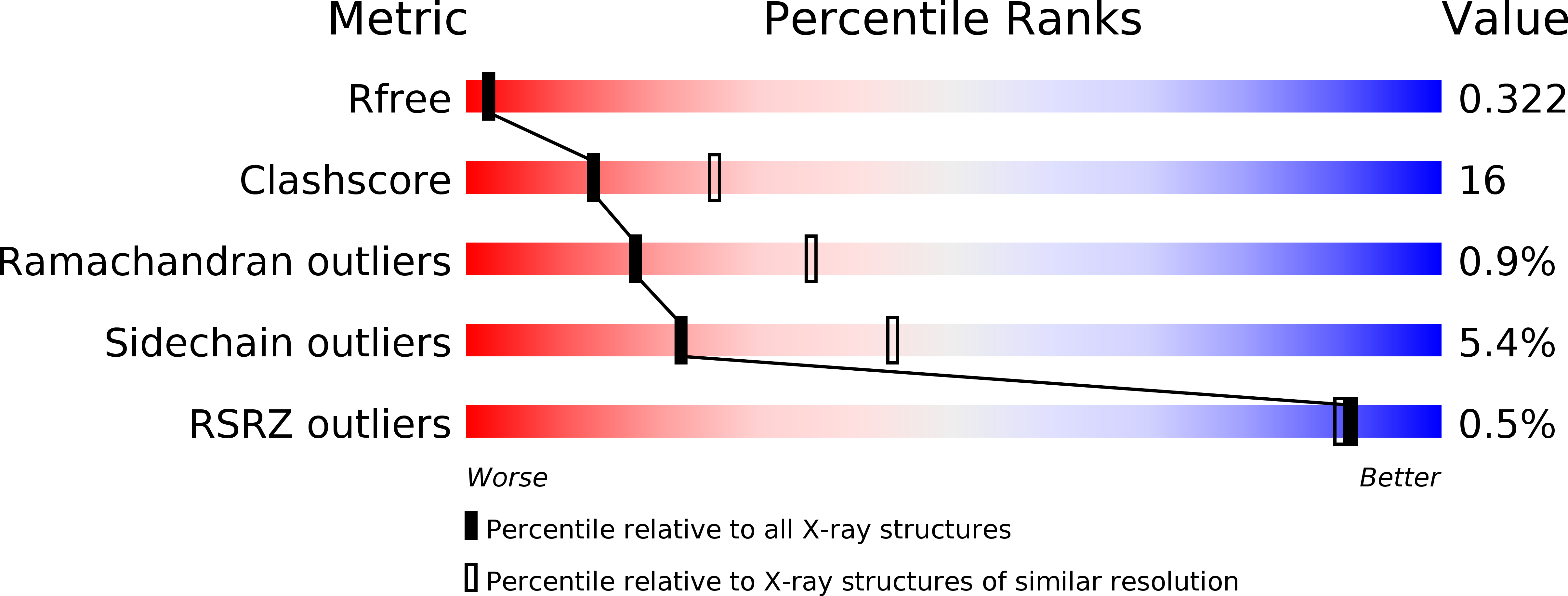
Resolution:
2.60 Å
R-Value Free:
0.32
R-Value Work:
0.22
R-Value Observed:
0.23
Space Group:
P 1 21 1
Macromolecular Entities
Polymer Type:polypeptide(L)
Molecule:ALPHA-1,4-GLUCAN LYASE ISOZYME 1
Gene (Uniprot):Ag111
Chain IDs:A, B, C, D
Chain Length:1027
Number of Molecules:4
Biological Source:GRACILARIOPSIS LEMANEIFORMIS
Modified Residue
Compound ID Chain ID Parent Comp ID Details 2D Image
CSO A CYS S-HYDROXYCYSTEINE
Ligand Molecules
Peptide-like Molecules
PRD_900022
Primary Citation
Crystal Structure of Alpha-1,4-Glucan Lyase, a Unique Glycoside Hydrolase Family Member with a Novel Catalytic Mechanism.
J.Biol.Chem. 288 26764 ? (2013)
PMID: 23902768 DOI: 10.1074/JBC.M113.485896

Abstact

α-1,4-Glucan lyase (EC 4.2.2.13) from the red seaweed Gracilariopsis lemaneiformis cleaves α-1,4-glucosidic linkages in glycogen, starch, and malto-oligosaccharides, yielding the keto-monosaccharide 1,5-anhydro-D-fructose. The enzyme belongs to glycoside hydrolase family 31 (GH31) but degrades starch via an elimination reaction instead of hydrolysis. The crystal structure shows that the enzyme, like GH31 hydrolases, contains a (β/α)8-barrel catalytic domain with B and B' subdomains, an N-terminal domain N, and the C-terminal domains C and D. The N-terminal domain N of the lyase was found to bind a trisaccharide. Complexes of the enzyme with acarbose and 1-dexoynojirimycin and two different covalent glycosyl-enzyme intermediates obtained with fluorinated sugar analogues show that, like GH31 hydrolases, the aspartic acid residues Asp(553) and Asp(665) are the catalytic nucleophile and acid, respectively. However, as a unique feature, the catalytic nucleophile is in a position to act also as a base that abstracts a proton from the C2 carbon atom of the covalently bound subsite -1 glucosyl residue, thus explaining the unique lyase activity of the enzyme. One Glu to Val mutation in the active site of the homologous α-glucosidase from Sulfolobus solfataricus resulted in a shift from hydrolytic to lyase activity, demonstrating that a subtle amino acid difference can promote lyase activity in a GH31 hydrolase.

Legend

Protein

Chemical

Disease

Primary Citation of related structures