2X2E image
Deposition Date 2010-01-12
Release Date 2010-04-28
Last Version Date 2024-11-13
Entry Detail
PDB ID:
2X2E
Keywords:
Title:
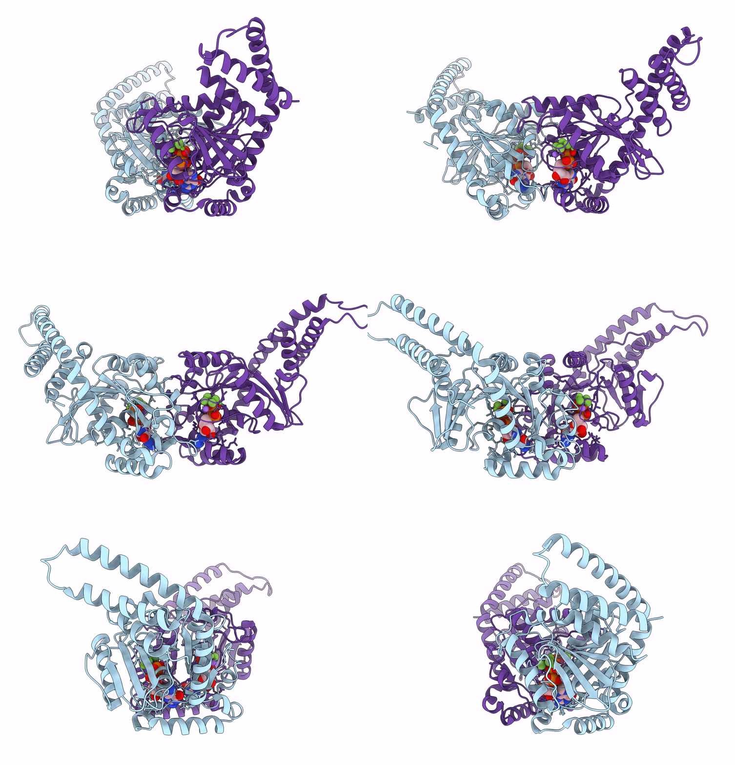
Dynamin GTPase dimer, long axis form
Biological Source:
Source Organism:
HOMO SAPIENS (Taxon ID: 9606)
Host Organism:
Method Details:
Experimental Method:
Resolution:
2.00 Å
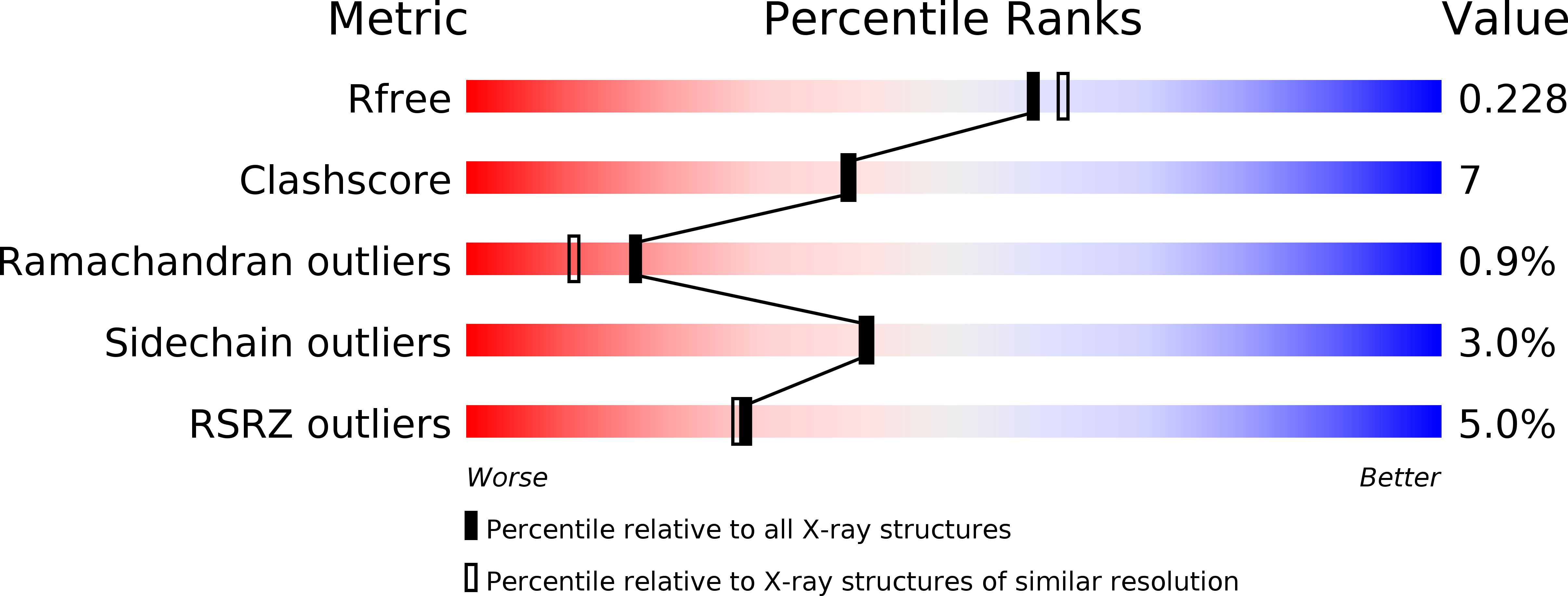
R-Value Free:
0.23
R-Value Work:
0.18
R-Value Observed:
0.18
Space Group:
P 21 21 21
Macromolecular Entities
Polymer Type:polypeptide(L)
Molecule:DYNAMIN-1
Gene (Uniprot):DNM1
Chain IDs:A, B (auth: D)
Chain Length:353
Number of Molecules:2
Biological Source:HOMO SAPIENS
Modified Residue
Compound ID Chain ID Parent Comp ID Details 2D Image
MSE A MET SELENOMETHIONINE
Primary Citation
G Domain Dimerization Controls Dynamin'S Assembly-Stimulated Gtpase Activity.
Nature 465 435 ? (2010)
PMID: 20428113 DOI: 10.1038/NATURE09032

Abstact

Dynamin is an atypical GTPase that catalyses membrane fission during clathrin-mediated endocytosis. The mechanisms of dynamin's basal and assembly-stimulated GTP hydrolysis are unknown, though both are indirectly influenced by the GTPase effector domain (GED). Here we present the 2.0 A resolution crystal structure of a human dynamin 1-derived minimal GTPase-GED fusion protein, which was dimeric in the presence of the transition state mimic GDP.AlF(4)(-).The structure reveals dynamin's catalytic machinery and explains how assembly-stimulated GTP hydrolysis is achieved through G domain dimerization. A sodium ion present in the active site suggests that dynamin uses a cation to compensate for the developing negative charge in the transition state in the absence of an arginine finger. Structural comparison to the rat dynamin G domain reveals key conformational changes that promote G domain dimerization and stimulated hydrolysis. The structure of the GTPase-GED fusion protein dimer provides insight into the mechanisms underlying dynamin-catalysed membrane fission.

Legend

Protein

Chemical

Disease

Primary Citation of related structures