
Deposition Date
2010-01-08
Release Date
2010-02-09
Last Version Date
2024-11-06
Entry Detail
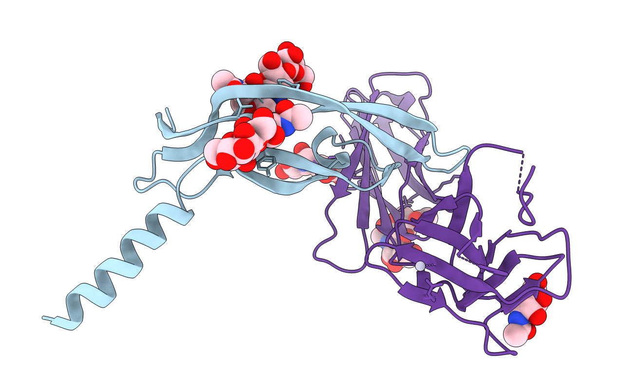
PDB ID:
2X1X
Keywords:
Title:
CRYSTAL STRUCTURE OF VEGF-C IN COMPLEX WITH DOMAINS 2 AND 3 OF VEGFR2 IN A TETRAGONAL CRYSTAL FORM
Biological Source:
Source Organism(s):
HOMO SAPIENS (Taxon ID: 9606)
Expression System(s):
Method Details:
Experimental Method:
Resolution:
3.10 Å
R-Value Free:
0.33
R-Value Work:
0.26
R-Value Observed:
0.27
Space Group:
P 42 21 2
