
Deposition Date
2009-12-17
Release Date
2009-12-29
Last Version Date
2023-12-20
Entry Detail
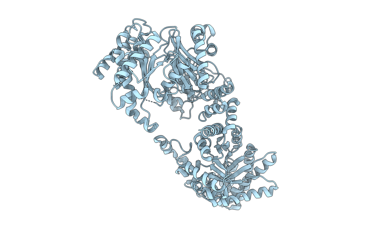
PDB ID:
2X0S
Keywords:
Title:
3.0 A RESOLUTION CRYSTAL STRUCTURE OF GLYCOSOMAL PYRUVATE PHOSPHATE DIKINASE FROM TRYPANOSOMA BRUCEI
Biological Source:
Source Organism(s):
TRYPANOSOMA BRUCEI (Taxon ID: 5691)
Expression System(s):
Method Details:
Experimental Method:
Resolution:
3.00 Å
R-Value Free:
0.24
R-Value Work:
0.16
R-Value Observed:
0.16
Space Group:
P 21 21 2
