Deposition Date
2009-11-30
Release Date
2009-12-15
Last Version Date
2024-05-08
Entry Detail
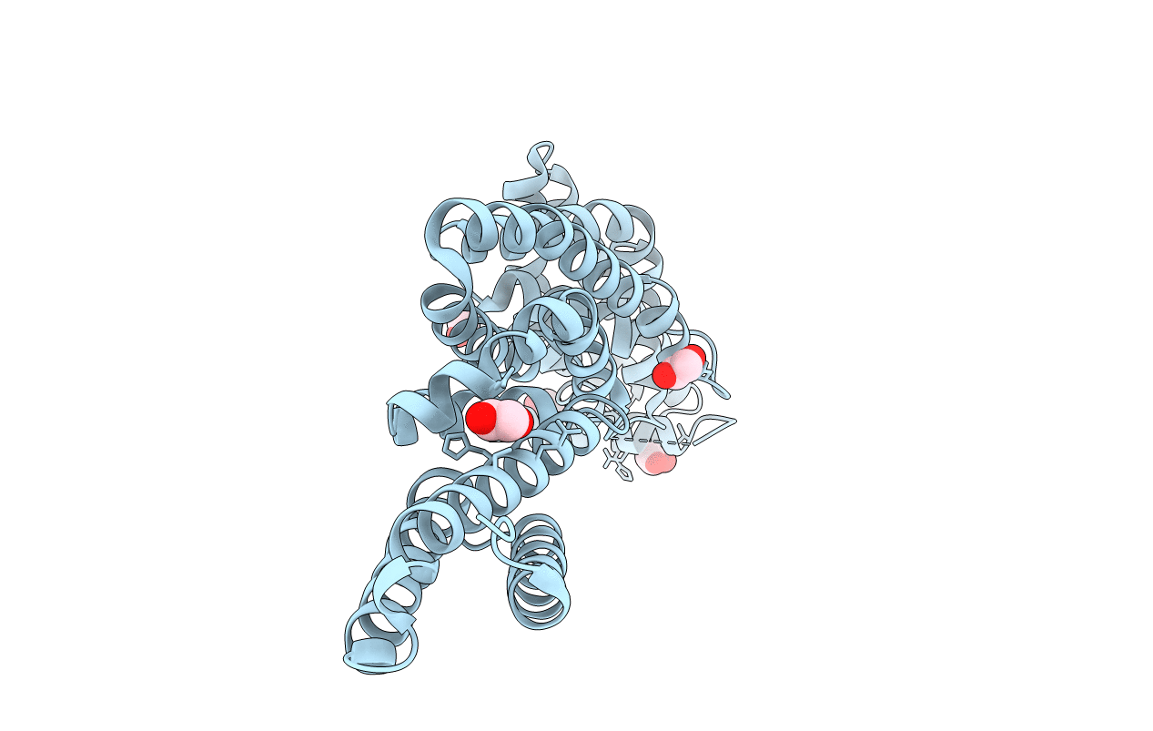
PDB ID:
2WZK
Keywords:
Title:
Structure of the Cul5 N-terminal domain at 2.05A resolution.
Biological Source:
Source Organism(s):
MUS MUSCULUS (Taxon ID: 10090)
Expression System(s):
Method Details:
Experimental Method:
Resolution:
2.05 Å
R-Value Free:
0.22
R-Value Work:
0.18
R-Value Observed:
0.19
Space Group:
P 21 21 21